

Massive loss of mid- and hindbrain neurons during embryonic development of homozygous lurcher mice
- PMID: 9065501
- PMCID: PMC6573489
- DOI: 10.1523/JNEUROSCI.17-07-02400.1997
Massive loss of mid- and hindbrain neurons during embryonic development of homozygous lurcher mice
Abstract
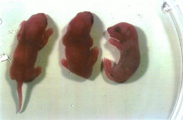
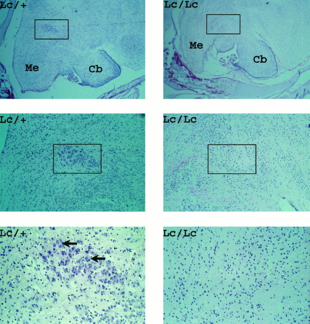
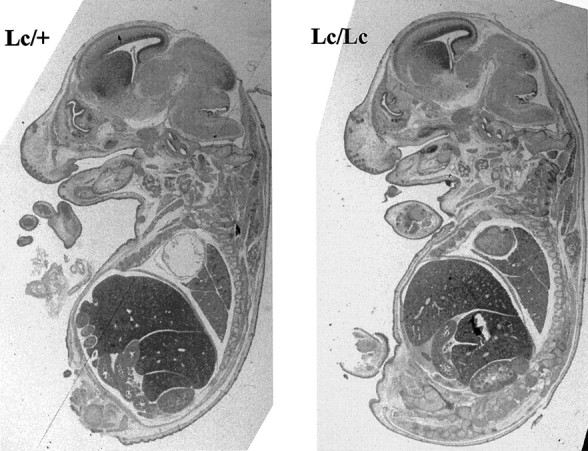
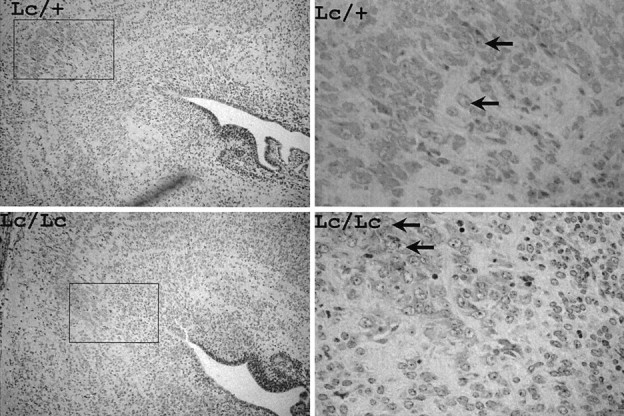
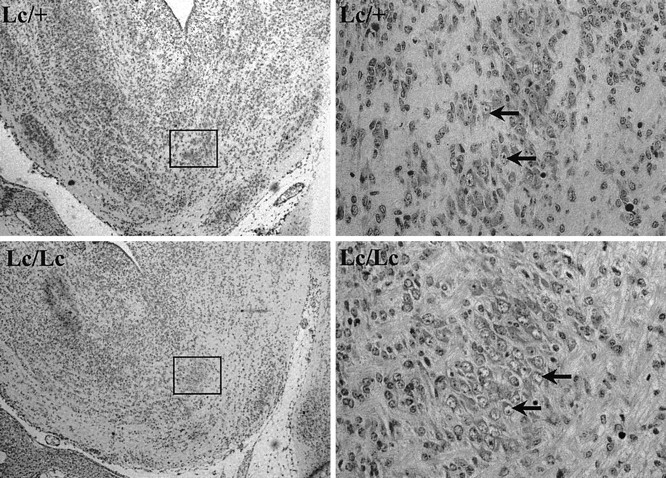
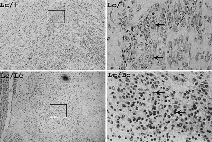
The mouse neurological mutant lurcher (Lc) results from a semidominant mutation. Heterozygous Lc/+ mice are viable but ataxic because Lc/+ Purkinje cells die by apoptosis within the first 3 weeks of life. Lc/Lc mice die shortly after birth. To aid in understanding the function of the lurcher gene product, we have examined the embryonic development of homozygous lurcher animals. The ratio of +/+:Lc/+:Lc/Lc animals did not deviate significantly from the expected 1:2:1. Homozygous lurcher mice at P0 were found to be normal under gross morphological examination. However, these mice weighed less, lacked milk in their stomach, and died within the first day of life. No resorbed embryos were found at embryonic day (E) 17.5, indicating that all homozygous lurchers survived until birth. Histological examination of P0 animals revealed that in homozygous lurcher mice the patterning of the brain is normal but that there has been a massive loss of hindbrain neurons during embryonic development. A particularly conspicuous consequence of the Lc/Lc genotype at birth is the complete absence of large neurons comprising the trigeminal motor nucleus. These neurons arise normally and are maintained until E15.5. However, beginning at E15.5 large numbers of pyknotic cells are evident in the trigeminal motor nucleus, suggesting that these cells die coincident with their terminal differentiation in the developing hindbrain. Because the trigeminal motor nucleus controls muscles required for suckling, these results suggest an explanation for the neonatal death of homozygous Lc animals. These data demonstrate that the severe and dose-dependent developmental consequences of lurcher gene action result from degeneration of distinct neuronal populations on maturation in the developing CNS.
Figures
References
-
- Barde YA. What, if anything, is a neurotrophic factor? Trends Neurosci. 1988;11:343–351. - PubMed
-
- Caddy KW, Briscoe TJ. Structural and quantitative studies on the normal C3H and lurcher mutant mouse. Philos Trans R Soc Lond [Biol] 1979;287:167–201. - PubMed
-
- Davies AM. Role of neurotrophic factors in development. Trends Genet. 1988;4:139–143. - PubMed
-
- Heckroth JA. Quantitative morphological analysis of the cerebellar nuclei in normal and lurcher mutant mice. I. Morphology and cell number. J Comp Neurol. 1994a;343:173–182. - PubMed
-
- Heckroth JA. A quantitative morphological analysis of the cerebellar nuclei in normal and lurcher mutant mice. II. Volumetric changes in cytological components. J Comp Neurol. 1994b;343:183–192. - PubMed
Publication types
MeSH terms
LinkOut - more resources
Full Text Sources
Molecular Biology Databases