

Neurogenesis in the dentate gyrus of the adult tree shrew is regulated by psychosocial stress and NMDA receptor activation
- PMID: 9065509
- PMCID: PMC6573503
- DOI: 10.1523/JNEUROSCI.17-07-02492.1997
Neurogenesis in the dentate gyrus of the adult tree shrew is regulated by psychosocial stress and NMDA receptor activation
Abstract
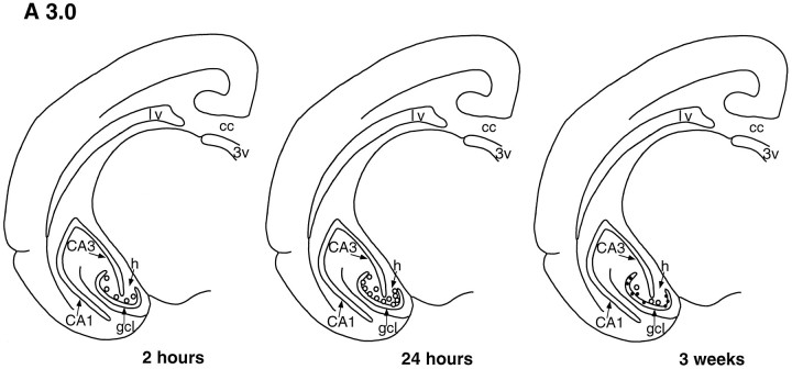
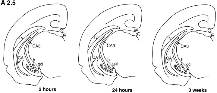
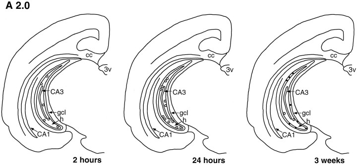
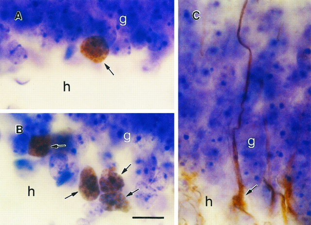
These studies were designed to determine whether adult neurogenesis occurs in the dentate gyrus of the tree shrew, an animal phylogenetically between insectivores and primates, and to explore the possibility that this process is regulated by stressful experiences and NMDA receptor activation. We performed immunohistochemistry for cell-specific markers and the thymidine analog bromodeoxyuridine (BrdU), a marker of DNA synthesis that labels proliferating cells and their progeny, on the brains of adult tree shrews subjected to psychosocial stress or NMDA receptor antagonist treatment. Cells that incorporated BrdU in the dentate gyrus of adult tree shrews were primarily located in the subgranular zone, had morphological characteristics of granule neuron precursors, and appeared to divide within 24 hr after BrdU injection. Three weeks after BrdU injection, BrdU-labeled cells had neuronal morphology, expressed the neuronal marker neuron specific enolase, and were incorporated into the granule cell layer. Vimentin-immunoreactive radial glia were observed in the dentate gyrus with cell bodies in the subgranular zone and processes extending into the granule cell layer. Exposure to acute psychosocial stress resulted in a rapid decrease in the number of BrdU-labeled cells in the dentate gyrus. In contrast, blockade of NMDA receptors, with the NMDA receptor antagonist MK-801, resulted in an increase in the number of BrdU-labeled cells in the dentate gyrus. These results indicate that adult neurogenesis occurs in the tree shrew dentate gyrus and is regulated by a stressful experience and NMDA receptor activation. Furthermore, we suggest that these characteristics may be common to most mammalian species.
Figures
References
-
- Altman J, Bayer SA. Mosaic organization of the hippocampal neuroepithelium and the multiple germinal sources of dentate granule cells. J Comp Neurol. 1990a;301:325–342. - PubMed
-
- Altman J, Bayer SA. Migration and distribution of two populations of hippocampal granule cell precursors during the perinatal and postnatal periods. J Comp Neurol. 1990b;301:365–381. - PubMed
-
- Altman J, Das GD. Postnatal neurogenesis in the guinea-pig. Nature. 1967;214:1098–1101. - PubMed
-
- Angevine JB. Time of neuron origin in the hippocampal region: an autoradiographic study in the mouse. Exp Neurol. 1965;2:1–17. - PubMed
-
- Bartanusz V, Aubry J-M, Pagliusi S, Jezova D, Baffi J, Kiss JZ. Stress-induced changes in messenger RNA levels of N-methyl-d-aspartate and AMPA receptor subunits in selected regions of the rat hippocampus and hypothalamus. Neuroscience. 1995;66:247–252. - PubMed
Publication types
MeSH terms
Substances
Grants and funding
LinkOut - more resources
Full Text Sources
Other Literature Sources
Medical