. 1997 Apr 1;17(7):2512-8.
doi: 10.1523/JNEUROSCI.17-07-02512.1997.

Environmental knowledge is subserved by separable dorsal/ventral neural areas

Affiliations

Environmental knowledge is subserved by separable dorsal/ventral neural areas

G K Aguirre et al. J Neurosci. .

Abstract

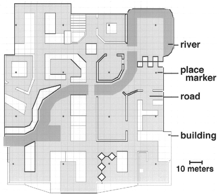
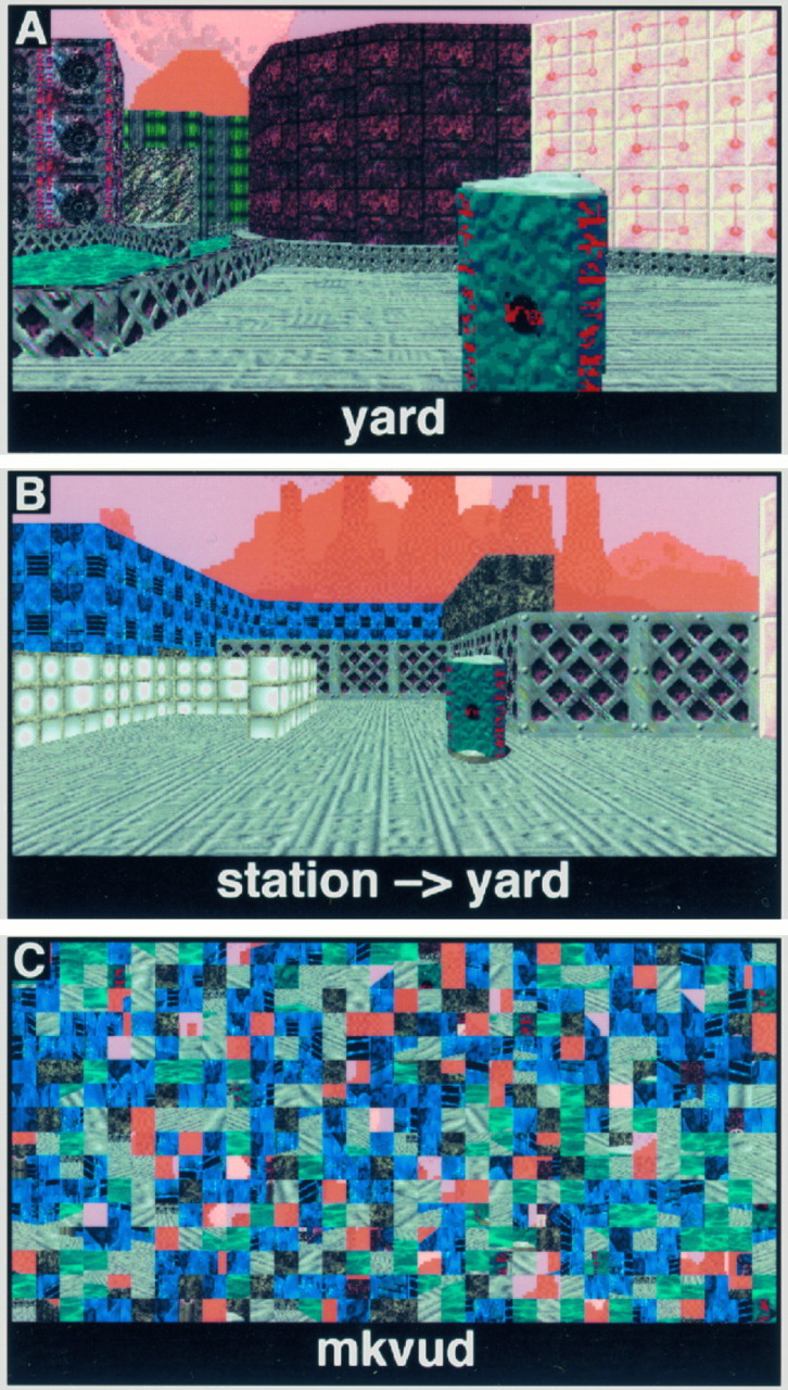
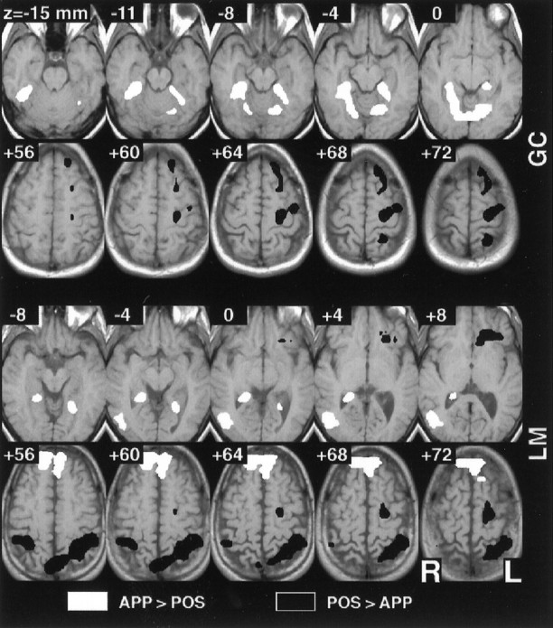
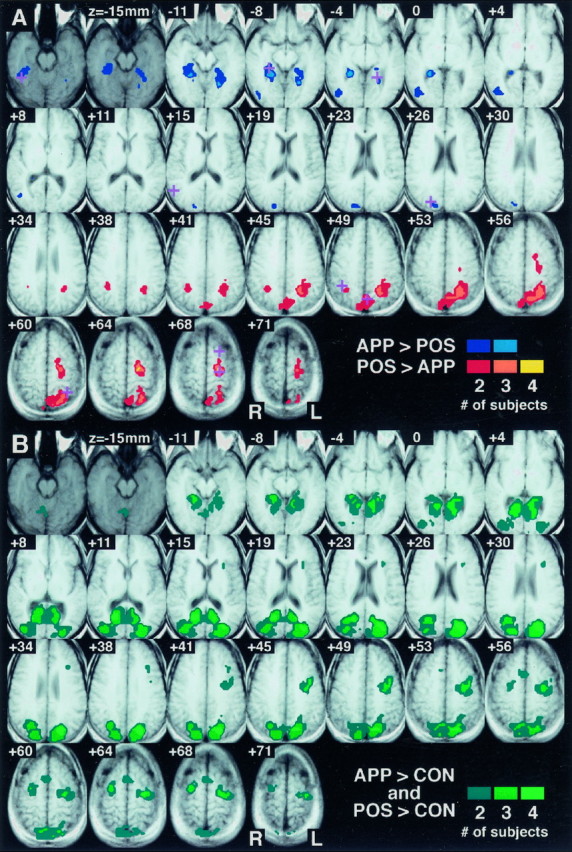
Environmental psychology models propose that knowledge of large-scale space is stored as distinct landmark (place appearance) and survey (place position) information. Studies of brain-damaged patients suffering from "topographical disorientation" tentatively support this proposal. In order to determine if the components of psychologically derived models of environmental representation are realized as distinct functional, neuroanatomical regions, a functional magnetic resonance imaging (fMRI) study of environmental knowledge was performed. During scanning, subjects made judgments regarding the appearance and position of familiar locations within a virtual reality environment. The fMRI data were analyzed in a manner that has been empirically demonstrated to rigorously control type I error and provide optimum sensitivity, allowing meaningful results in the single subject. A direct comparison of the survey position and landmark appearance conditions revealed a dorsal/ventral dissociation in three of four subjects. These results are discussed in the context of the observed forms of topographical disorientation and are found to be in good agreement with the human lesion studies. This experiment confirms that environmental knowledge is not represented by a unitary system but is instead functionally distributed across the neocortex.


Figures

Fig. 1.
Fig. 1.
Aerial view of the virtual reality town. The town contained 16 distinctive, named “places” interconnected by a variety of roads and paths and arranged in a 4 × 4 grid. A river divided the town approximately in half. Subjectively, the town was ∼140 meters in width. Each place was designated as such by the presence of a small round marker in the ground.
Fig. 2.
Fig. 2.
Behavioral task stimuli. A, APPEARANCE task; the subject indicated whether the name matched the pictured place. B, POSITION task; the identity of the subject’s current location was given, and the subject was asked to indicate the direction of a second, target place. All targets were located in a cardinal direction (forward, behind, left or right). Note that the “sky” background, although textured, did not provide any useful orienting information. C, CONTROL task; different scrambled words and scenes were presented in response to the subject alternating left and right button presses.
Fig. 3.
Fig. 3.
Selected individual subject results for the direct POSITION–APPEARANCE comparison. Individual subject data were analyzed using the modified general linear model for autocorrelated observations and the resulting t maps thresholded atα = 0.025 (corrected for multiple comparisons). The maps have been converted to standard Talairach space for ease of comparison, and four adjacent inferior and superior axial slices are depicted for each of two subjects. Greater activity was seen in the inferior and superior parietal lobule and the premotor cortex during the POSITION task, whereas greater activity was observed in the parahippocampal, lingual, and fusiform gyri during the APPEARANCE task.
Fig. 4.
Fig. 4.
Sites of replicated significant activity across four subjects. Shown are 25 axial slices, spaced every 3.75 mm, through the observed volume. Data are displayed on averaged T1 localizers from the four subjects converted to standard Talairach space. The most inferior two axial slices are presented in a darker graythan the others to indicate that this inferior level was imaged for only two of the four subjects. As a result, the maximum overlap possible for these slices is two. Points where multiple subjects possessed significant activity are indicated in color.A, Direct POSITION–APPEARANCE comparison; the plus symbols indicate the sites of clusters of local maxima present in multiple individual subjects and correspond to the locations listed in Table 1. B, POSITION versus CONTROL and APPEARANCE versus CONTROL comparisons; shown are those points where multiple subjects had greater activity during both the APPEARANCE and the POSITION tasks as compared with CONTROL.

Similar articles

Cited by

References

    1. Aguirre GK, Detre JA, Alsop DC, D’Esposito M. The parahippocampus subserves topographical learning in man. Cereb Cortex. 1996;6:823–829. - PubMed
    1. Aguirre GK, Zarahn E, D’Esposito M (1997) Empirical analyses of BOLD fMRI statistics. II. Spatially smoothed data collected under null-hypothesis and experimental conditions. NeuroImage, in press. - PubMed
    1. Allison T, Ginter H, McCarthy G, Nobre AC, Puce A, Luby M, Spencer DD. Face recognition in human extrastriate cortex. J Neurophysiol. 1994;71:821–825. - PubMed
    1. Bottini G, Cappa S, Geminiani G, Sterzi R. Topographic disorientation: a case report. Neuropsychologia. 1990;28:309–312. - PubMed
    1. Cohen JD, MacWhinney M, Flatt M, Provost J. PsyScope: a new graphic interactive environment for designing psychology experiments. Behav Res Methods Instrum Comput. 1993;25:257–271.

Publication types