. 1997 Mar 24;136(6):1185-99.
doi: 10.1083/jcb.136.6.1185.

In vivo dynamics of nuclear pore complexes in yeast

Affiliations

In vivo dynamics of nuclear pore complexes in yeast

M Bucci et al. J Cell Biol. .

Abstract

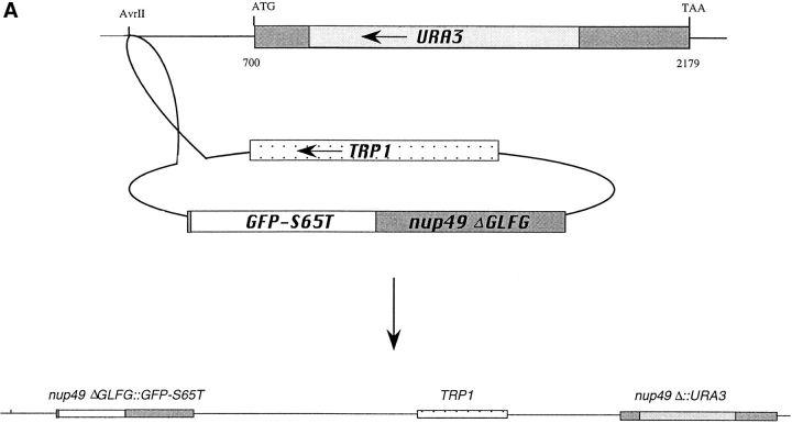
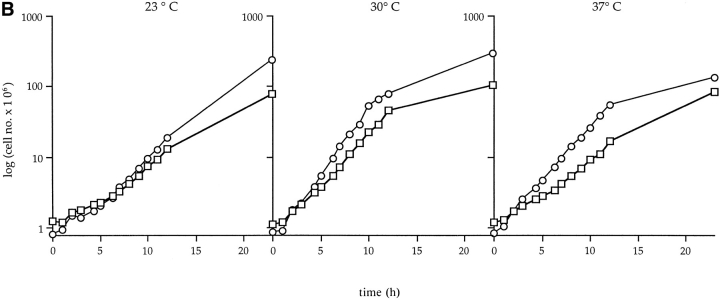
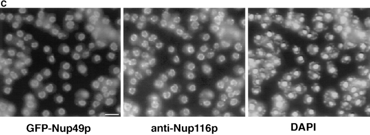
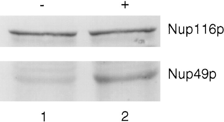
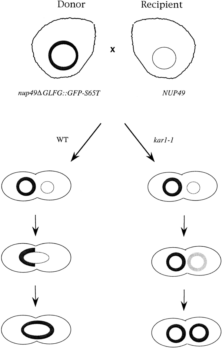
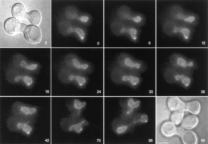
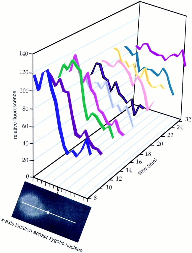
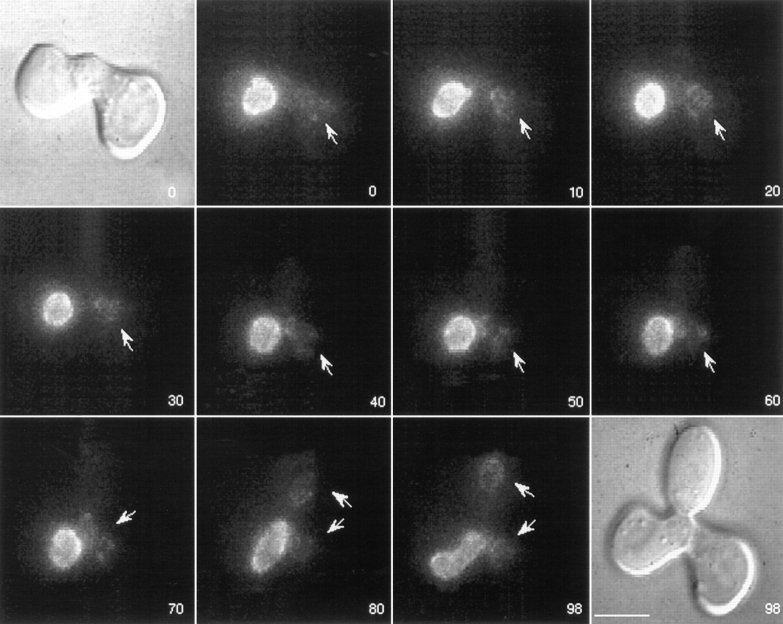
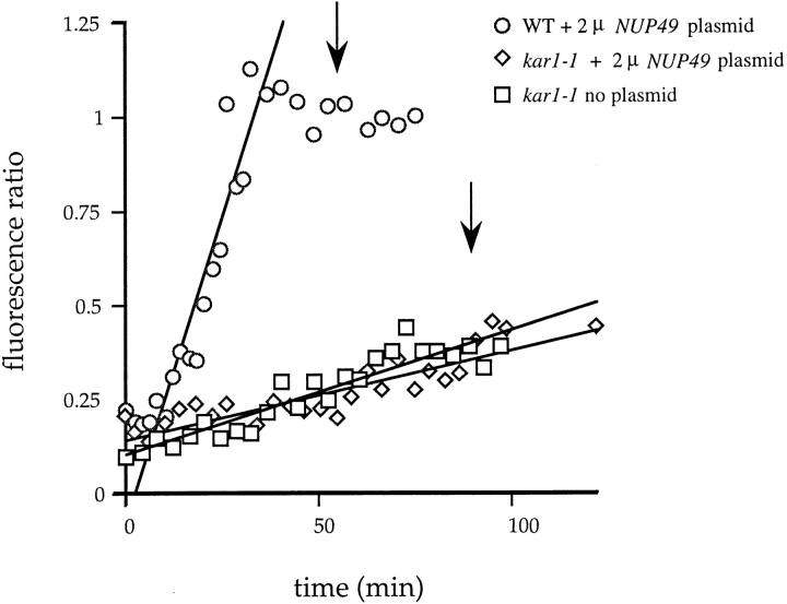
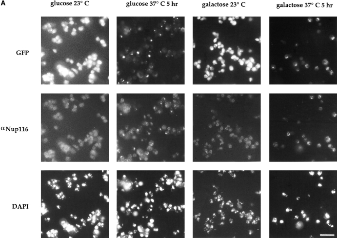
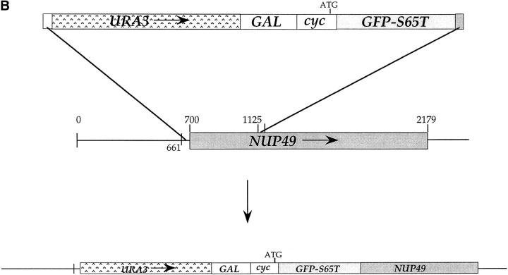
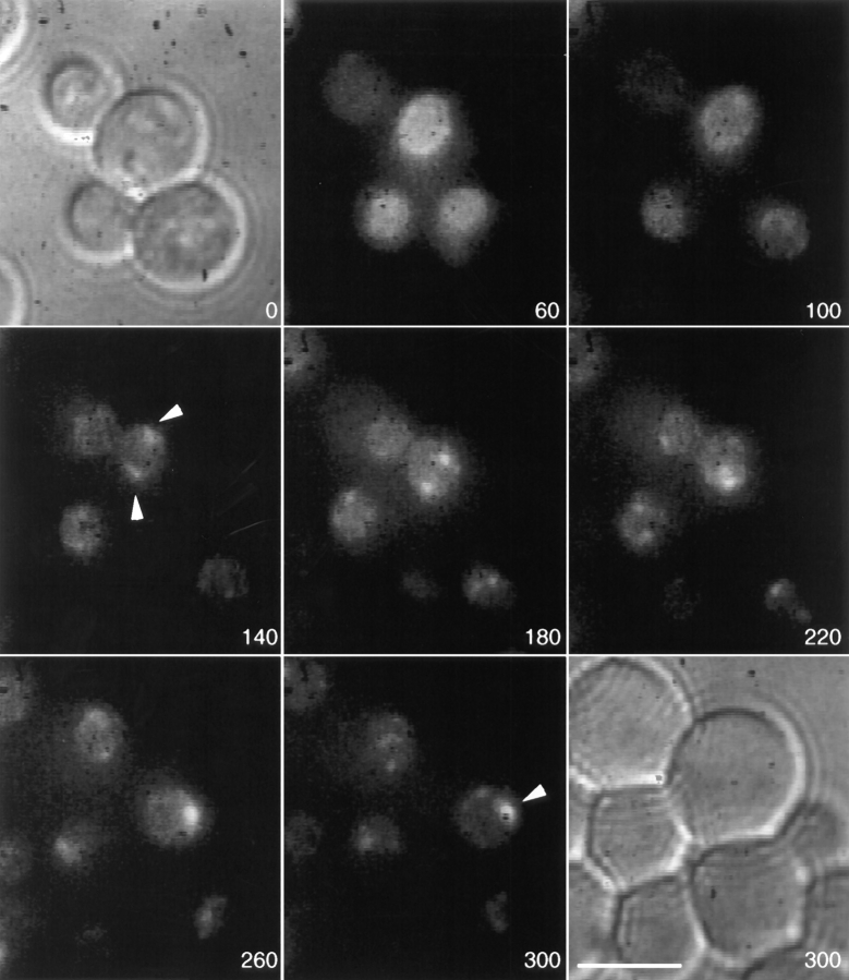
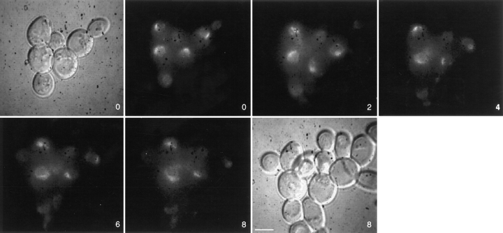
While much is known about the role of nuclear pore complexes (NPCs) in nucleocytoplasmic transport, the mechanism of NPC assembly into pores formed through the double lipid bilayer of the nuclear envelope is not well defined. To investigate the dynamics of NPCs, we developed a live-cell assay in the yeast Saccharomyces cerevisiae. The nucleoporin Nup49p was fused to the green fluorescent protein (GFP) of Aequorea victoria and expressed in nup49 null haploid yeast cells. When the GFP-Nup49p donor cell was mated with a recipient cell harboring only unlabeled Nup49p, the nuclei fused as a consequence of the normal mating process. By monitoring the distribution of the GFP-Nup49p, we could assess whether NPCs were able to move from the donor section of the nuclear envelope to that of the recipient nucleus. We observed that fluorescent NPCs moved and encircled the entire nucleus within 25 min after fusion. When assays were done in mutant kar1-1 strains, where nuclear fusion does not occur, GFP-Nup49p appearance in the recipient nucleus occurred at a very slow rate, presumably due to new NPC biogenesis or to exchange of GFP-Nup49p into existing recipient NPCs. Interestingly, in a number of existing mutant strains, NPCs are clustered together at permissive growth temperatures. This has been explained with two different hypotheses: by movement of NPCs through the double nuclear membranes with subsequent clustering at a central location; or, alternatively, by assembly of all NPCs at a central location (such as the spindle pole body) with NPCs in mutant cells unable to move away from this point. Using the GFP-Nup49p system with a mutant in the NPC-associated factor Gle2p that exhibits formation of NPC clusters only at 37 degrees C, it was possible to distinguish between these two models for NPC dynamics. GFP-Nup49p-labeled NPCs, assembled at 23 degrees C, moved into clusters when the cells were shifted to growth at 37 degrees C. These results indicate that NPCs can move through the double nuclear membranes and, moreover, can do so to form NPC clusters in mutant strains. Such clusters may result by releasing NPCs from a nuclear tether, or by disappearance of a protein that normally prevents pore aggregation. This system represents a novel approach for identifying regulators of NPC assembly and movement in the future.


Figures

Figure 2
Figure 2
GFP–Nup49p is functional and localizes to NPCs. (A) Plasmid DNA encoding for GFP–Nup49p fusion protein was cut with AvrII for chromosomal integration into a diploid strain heterozygous for the NUP49 null allele, nup49Δ::URA3. Haploids expressing GFP– Nup49p were obtained by sporulation and dissection of Trp+ Ura+ diploids in which TRP1 and URA3 markers cosegregated. (B) Cells expressing GFP–Nup49p grow at rates comparable to wildtype cells at all temperatures. Growth curves are plotted as the log10 of the cell concentration over time. (Circles) SWY809, nup49ΔGLFG:: GFP–S65T cells; (squares) SWY518, wild-type cells. (C) GFP–Nup49p colocalizes with the nucleoporin Nup116p. Immunofluorescence was performed on cells expressing GFP–Nup49p using an affinity-purified polyclonal antibody against Nup116p. GFP fluorescence was visualized directly, and the antiNup116p antibody was visualized using a Texas red– conjugated goat anti–rabbit antibody. Nuclear DNA was visualized by DAPI staining. Bar, 10 μm.
Figure 2
Figure 2
GFP–Nup49p is functional and localizes to NPCs. (A) Plasmid DNA encoding for GFP–Nup49p fusion protein was cut with AvrII for chromosomal integration into a diploid strain heterozygous for the NUP49 null allele, nup49Δ::URA3. Haploids expressing GFP– Nup49p were obtained by sporulation and dissection of Trp+ Ura+ diploids in which TRP1 and URA3 markers cosegregated. (B) Cells expressing GFP–Nup49p grow at rates comparable to wildtype cells at all temperatures. Growth curves are plotted as the log10 of the cell concentration over time. (Circles) SWY809, nup49ΔGLFG:: GFP–S65T cells; (squares) SWY518, wild-type cells. (C) GFP–Nup49p colocalizes with the nucleoporin Nup116p. Immunofluorescence was performed on cells expressing GFP–Nup49p using an affinity-purified polyclonal antibody against Nup116p. GFP fluorescence was visualized directly, and the antiNup116p antibody was visualized using a Texas red– conjugated goat anti–rabbit antibody. Nuclear DNA was visualized by DAPI staining. Bar, 10 μm.
Figure 7
Figure 7
Cells harboring the 2 μ NUP49 plasmid have elevated levels of Nup49p. Yeast strains SWY459 (wild type, lane 1) and SWY759 (SWY459 cells harboring the 2 μ NUP49 plasmid, lane 2) were grown in synthetic media lacking uracil or tryptophan, respectively. Total protein extracts were prepared from equivalent numbers of cells, separated on a 9% SDS polyacrylamide gel, and transferred to nitrocellulose. Blots were probed with an affinitypurified polyclonal antibody raised against the GLFG region of Nup116p that also recognizes Nup49p (gift of K. Iovine and J. Watkins, Washington University, St. Louis, MO).
Figure 1
Figure 1
NPC movement and assembly assay. A haploid yeast strain expressing only GFP–Nup49p (Donor) is mated with a haploid strain expressing unlabeled Nup49p (Recipient). In the case where both strains are otherwise wild type (WT), the labeled nucleus (thick circle) will fuse with the recipient nucleus (thin circle) upon mating. Movement of NPCs can then be monitored by watching GFP–Nup49p redistribution in live cells. In a kar1-1 background (kar1-1), the nuclei are unable to fuse and GFP- labeled NPCs are obtained by the recipient nucleus only by incorporation into preexisting/new NPCs. The rate of acquisition of GFP–NPCs in the recipient nuclei reflects either the movement of NPCs or the assembly of NPCs, respectively.
Figure 3
Figure 3
Wild-type NPCs move within the nuclear envelope. Cells expressing GFP–Nup49p (SWY809) were mixed with cells of the opposite mating type expressing wild-type Nup49p from the chromosome and a high copy number plasmid (SWY759). After 5 h in culture, cells were prepared for video microscopy by incubating mating mixtures on an agarose-covered slide. Both Nomarski and fluorescence images for the 0- and 98-min time points are shown. Two newly formed zygotes are shown. GFP–Nup49p distribution was recorded every 2 min for 98 min, and selected frames are shown here. Each fluorescent image is a two-dimensional projection of all z-axis planes. By the end of the sequence (98 min panel), the two zygotes are at various stages of nuclear division, allowing nuclear segregation into the newly formed daughter buds (Nomarski image, 98 min). Numbers indicate time in min. Bar, 5 μm.
Figure 4
Figure 4
Time course of GFP–Nup49p redistribution in the zygotic nucleus. A representative image is shown from early in the video of the top wild-type zygote in Fig. 3. At each time point during the video (y-axis), a line spanning the donor and recipient nuclear surfaces was designated (shown in white with the dot marking the fusion junction). Distance along the line is graphed on the x-axis. For each line in the respective zygotic nucleus, NIH Image 1.60 was used to quantify the fluorescence intensity at 28 points ∼0.3 μm apart. The fluorescence values of each two sequential measurements along the x-axis were averaged, and the 14 resulting relative fluorescence data points were graphed on the z-axis. The first time point at 8 min in the video (blue ribbon) represents the approximate time of nuclear fusion. Later time points are shown in different colors along the y-axis. Microsoft Excel 4.0 (Seattle, WA) was used to display the graph.
Figure 5
Figure 5
NPC assembly follows a slower time course than NPC movement. kar1-1 mutant cells expressing GFP–Nup49p (SWY1308) were mixed with cells of the opposite mating type expressing both genomic unlabeled Nup49p and unlabeled Nup49p from a high copy 2 μ plasmid (SWY757). GFP–NPC distribution was recorded in two-dimensional projection videos as described in Fig. 3. Selected frames from a 98-min video are shown. The first and last frames are Nomarski images taken at the beginning and end of the video: note that the two nuclei in the zygote have not fused. The recipient nucleus (arrows) slowly acquired GFP fluorescence but divided before it reached the intensity of the GFP–Nup49p donor nucleus. Numbers indicate time in min. Bar, 5 μm.
Figure 6
Figure 6
NPC movement rates are distinct from GFP–Nup49p incorporation rates. Quantification of NPC movement and assembly was conducted using NIH Image to calculate the average pixel brightness value of an area encircling the donor or recipient nucleus. The fluorescence ratio was determined by dividing the average brightness value of the recipient nucleus by the average brightness value of the donor nucleus. The data for one zygote for each type of mating are shown. (Circles) Wild-type zygote with 2 μ NUP49 plasmid; (diamonds) kar1-1 zygote with 2 μ NUP49 plasmid; (squares) kar1-1 zygote with no plasmid. The Cricket Graph program was used to fit a line through the linear portion of the wild-type data set, and through all the points for the kar1-1 experiments. (Arrows) Approximate time of nuclear division for wild-type (top arrow) and kar1-1 zygotes (bottom arrow).
Figure 8
Figure 8
NPCs move into clusters in the gle2-1 temperature-sensitive mutant. (A) gle2-1 cells expressing GFP– Nup49p under control of the galactose-inducible promoter and Nup49p from a 2 μ plasmid (SWY1324) were grown in synthetic minimal medium lacking histidine and containing 0.5% galactose and 1.5% raffinose. Half of the cells were washed out of galactose and into glucosecontaining media for 1 h before a shift to the nonpermissive temperature of 37°C. Cells were maintained at 37°C in glucose or galactose for 5 h before processing for direct visualization and immunofluorescence as described for Fig. 2 C. Clusters of NPCs appear in cells shifted to glucose as well as those maintained in galactose. (B) PCR was performed on pSW441 to create a fragment flanked by sequences complementary to NUP49. The fragment was transformed into wild-type SWY595 cells for integration onto the chromosome. Bar, 10 μm.
Figure 8
Figure 8
NPCs move into clusters in the gle2-1 temperature-sensitive mutant. (A) gle2-1 cells expressing GFP– Nup49p under control of the galactose-inducible promoter and Nup49p from a 2 μ plasmid (SWY1324) were grown in synthetic minimal medium lacking histidine and containing 0.5% galactose and 1.5% raffinose. Half of the cells were washed out of galactose and into glucosecontaining media for 1 h before a shift to the nonpermissive temperature of 37°C. Cells were maintained at 37°C in glucose or galactose for 5 h before processing for direct visualization and immunofluorescence as described for Fig. 2 C. Clusters of NPCs appear in cells shifted to glucose as well as those maintained in galactose. (B) PCR was performed on pSW441 to create a fragment flanked by sequences complementary to NUP49. The fragment was transformed into wild-type SWY595 cells for integration onto the chromosome. Bar, 10 μm.
Figure 9
Figure 9
Monitoring movement of NPCs into clusters. SWY1324 cells were grown as in Fig. 8 A to induce expression of GFP–Nup49p. Cells were then washed into media containing 2% glucose for 5 h. Cells were shifted to 37°C on a slide set on a temperature-controlled microscope stage. Videos were taken with time points every 20 min for 5 h as described for Fig. 3. Selected frames 40 min apart are shown. The first and last frames are Nomarski images before and after the video. Numbers indicate time after the 37°C shift in min. (Arrowheads) NPC clusters formed by NPC movement. Bar, 5 μm.
Figure 10
Figure 10
Cluster formation in gle2-1 cells is not rapidly reversed. SWY1324 cells were induced at 23°C to express GFP–Nup49p by growing in SC − his containing 0.5% galactose/1.5% raffinose. Cells were then shifted to 37°C in SC − his with 2% glucose for 5 h before a return to 23°C. Videos were taken as described for the movement assay in Fig. 3. Frames were taken every 30 min for 8 h after returning the cells to 23°C. Both Nomarski and fluorescence images were taken at the 0- and 8-h time points. Selected frames are shown here. Bar, 5 μm.

References

    1. Aitchison JD, Blobel G, Rout MP. Nup120p: a yeast nucleoporin required for NPC distribution and mRNA transport. J Cell Biol. 1995a;131:1659–1675. - PMC - PubMed
    1. Aitchison JD, Rout MP, Marelli M, Blobel G, Wozniak RW. Two novel related yeast nucleoporins Nup170p and Nup157p : complementation with the vertebrate homologue Nup155p and functional interactions with the yeast nuclear pore–membrane protein Pom152p. J Cell Biol. 1995b;131:1133–1148. - PMC - PubMed
    1. Akey CW. Structural plasticity of the nuclear pore complex. J Mol Biol. 1995;248:273–293. - PubMed
    1. Aris JP, Blobel G. Yeast nuclear envelope proteins cross-react with an antibody against mammalian pore complex proteins. J Cell Biol. 1989;108:2059–2067. - PMC - PubMed
    1. Baudin A, Ozier KO, Denouel A, Lacroute F, Cullin C. A simple and efficient method for direct gene deletion in Saccharomyces cerevisiae. . Nucleic Acids Res. 1993;21:3329–3330. - PMC - PubMed

Publication types

MeSH terms