. 1997 Mar 24;136(6):1213-26.
doi: 10.1083/jcb.136.6.1213.

Identification of a novel stage of ribosome/nascent chain association with the endoplasmic reticulum membrane

Affiliations

Identification of a novel stage of ribosome/nascent chain association with the endoplasmic reticulum membrane

E C Murphy 3rd et al. J Cell Biol. .

Abstract

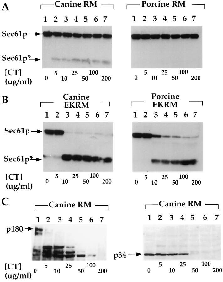
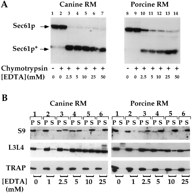
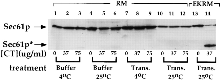
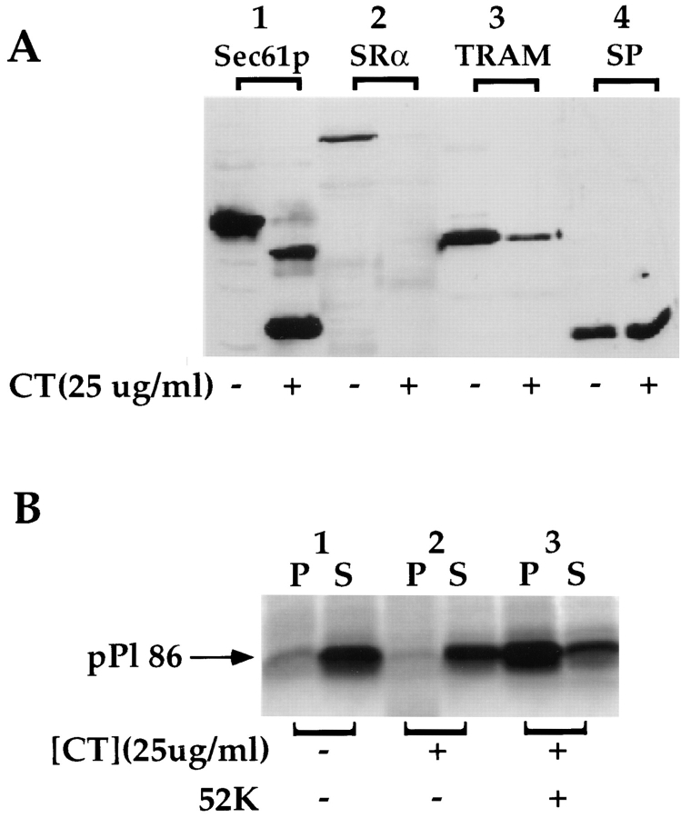
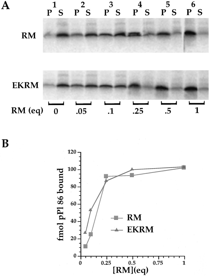
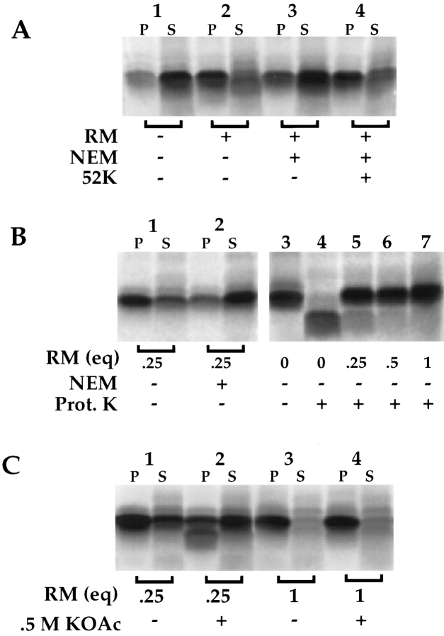
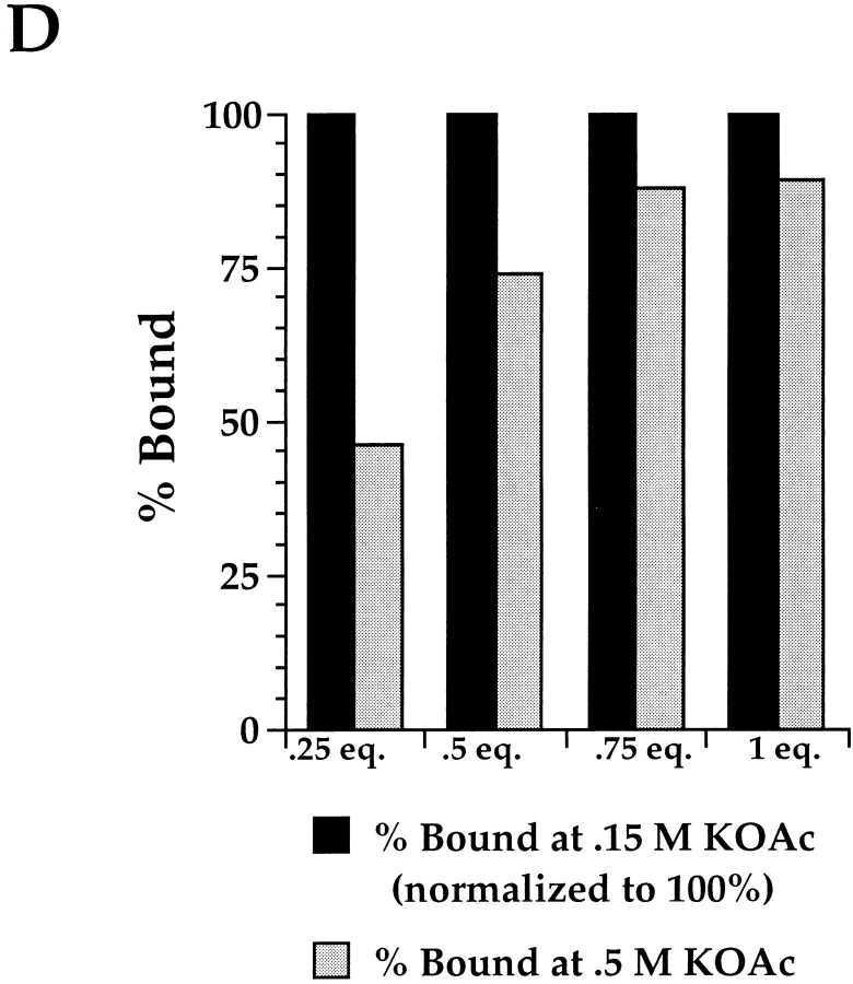
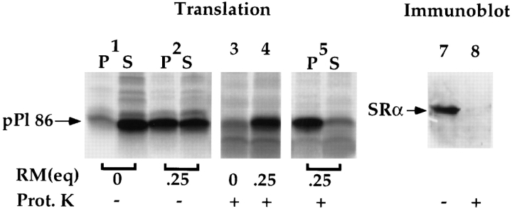
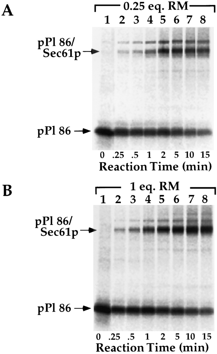
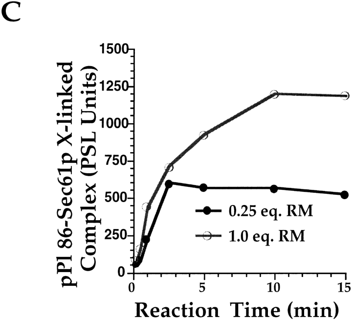
Protein translocation in the mammalian endoplasmic reticulum (ER) occurs cotranslationally and requires the binding of translationally active ribosomes to components of the ER membrane. Three candidate ribosome receptors, p180, p34, and Sec61p, have been identified in binding studies with inactive ribosomes, suggesting that ribosome binding is mediated through a receptor-ligand interaction. To determine if the binding of nascent chain-bearing ribosomes is regulated in a manner similar to inactive ribosomes, we have investigated the ribosome/nascent chain binding event that accompanies targeting. In agreement with previous reports, indicating that Sec61p displays the majority of the ER ribosome binding activity, we observed that Sec61p is shielded from proteolytic digestion by native, bound ribosomes. The binding of active, nascent chain bearing ribosomes to the ER membrane is, however, insensitive to the ribosome occupancy state of Sec61p. To determine if additional, Sec61p independent, stages of the ribosome binding reaction could be identified, ribosome/nascent chain binding was assayed as a function of RM concentration. At limiting RM concentrations, a protease resistant ribosome-membrane junction was formed, yet the nascent chain was salt extractable and cross-linked to Sec61p with low efficiency. At nonlimiting RM concentrations, bound nascent chains were protease and salt resistant and cross-linked to Sec61p with higher efficiency. On the basis of these and other data, we propose that ribosome binding to the ER membrane is a multi-stage process comprised of an initial, Sec61p independent binding event, which precedes association of the ribosome/nascent chain complex with Sec61p.


Figures

Figure 1
Figure 1
Protease protection of ER proteins in RM and EKRM. 4 eq. of either RM (A and C) or EKRM (B) were diluted to 20 μl in a buffer containing 25 mM K-Hepes, pH 7.2, 25 mM KOAc, and 2.5 mM Mg(OAc)2 at 4°C. Samples were treated with chymotrypsin (CT) at the indicated concentration for 30 min at 4°C and reactions quenched by addition of TCA to 10%. After centrifugation, samples were processed for SDS-PAGE and immunoblotted with antibodies directed against Sec61p, p180, and p34, as described in Materials and Methods. The migration of full-length Sec61p, p180, and p34 are indicated by arrows; a prominent limit digestion product of Sec61p is indicated by an asterisk.
Figure 2
Figure 2
EDTA treatment of RM: effects on Sec61p susceptibility to protease digestion and release of bound 60 and 40 S ribosomal subunits. 4 eq. of either canine or porcine RM were diluted to 20 μl in buffer containing 25 mM K-Hepes pH 7.2, 25 mM KOAc at 4°C. After dilution, samples were treated with EDTA at the indicated concentration for 15 min at 4°C, and either treated with chymotrypsin (25 μg/ml) for 30 min at 4°C (A), or centrifuged to separate membraneassociated and free ribosomal subunits (B). (A) After chymotrypsin treatment and acid precipitation, samples were processed for SDSPAGE and immunoblotted with an antibody directed against Sec61p. As in Fig. 1, the migration of both full- length Sec61p and the limit digestion product are indicated. (B) After EDTA treatment, samples were diluted sevenfold in a physiological salts buffer, layered over a 0.5-M sucrose cushion, and centrifuged to separate bound from free ribosomal subunits (6 min, 60,000 rpm, TLA 100 rotor, 4°C). Pellet (P) and supernatant (S) samples were processed for SDSPAGE, and immunoblotted with antibodies directed against the 40-S subunit protein S9, 60 S subunit proteins L3 and L4 (L3L4), or TRAPα.
Figure 3
Figure 3
Run-off translation does not alter the accessibility of Sec61p to proteolytic digestion. RM were treated with buffer (25 mM K-Hepes, pH 7.2, 110 mM KOAc, 2.5 mM Mg(OAc)2) at 4°C or 25°C, or with reticulocyte translation mixture (see Materials and Methods) at 4°C or 2°C. Subsequently, samples were treated with the indicated concentration of chymotrypsin for 30 min at 4°C. Samples treated with buffer were processed for SDS-PAGE as described previously. Samples treated with reticulocyte translation mixture were diluted sevenfold in physiological salt buffer, overlaid onto a 0.5-M sucrose cushion, and centrifuged for 10 min at 60,000 rpm in a TLA 100 rotor, as described in the legend to Fig. 2. The pellet (RM) fraction was solubilized in SDS-PAGE sample buffer and processed for SDS-PAGE. After transfer to nitrocellulose, samples were immunoblotted with an antibody directed against Sec61p.
Figure 4
Figure 4
Inactive ribososomes do not compete for binding with ribosome/nascent chain complexes. pPl 86 was translated in the absence of RM, and aliquots of the translation reaction (20 μl) added to 1 eq. of RM, subsequent to addition of the indicated quantity of 80 S reticulocyte lysate-derived ribosomes. Buffer conditions were adjusted such that the final KOAc concentration was 140 mM. After incubation at 25°C for 10 min, RM-bound pPl 86 was separated from unbound by centrifugation through a 0.5-M sucrose cushion as described in the legend to Fig. 2, and samples processed for SDS-PAGE. Quantitation of the [35S] pPl 86 was performed by phosphorimager analysis of the dried gels on a Fuji MacBAS 1000 phosphorimager.
Figure 5
Figure 5
Proteolysis of Sec61p does not alter ribosome/pPl 86 binding. (A) Untreated and chymotrypsin-treated EKRM were resolved on 12.5% SDS-PAGE gels, and immunoblotted with antibodies directed against Sec61p, SRα, TRAM, and the 22/23-kD subunit of the signal peptidase complex. (B) EKRM were treated with chymotrypsin at 25 μg/ml for 30 min at 4°C, conditions known to yield nearly quantitative digestion of Sec61p. After protease treatment, chymotrypsin-treated EKRM were tested for their ability to support binding of ribosome/pPl 86, either in the presence or absence of the 52-kD fragment of SRα. pPl 86 was translated in the absence of 1.0 eq. EKRM (lane 1), in the presence of 1.0 eq. chymotrypsin-treated EKRM (lane 2), and in the presence of 1.0 eq. chymotrypsin-treated EKRM supplemented with 52 kD SRα fragment (lane 3). The 52-kD SRα fragment was incubated with chymotrypsin-treated EKRM for 30 min at 4°C, before translation.
Figure 6
Figure 6
Ribosome/pPl 86 binding capacity of RM and EKRM. pPl 86 was translated in the absence of microsomes and, following translation, 20-μl aliquots of the translation were added to the indicated quantities of either RM or EKRM and a binding reaction performed for 10 min at 25°C. Samples were diluted sevenfold using a buffer containing 25 mM K-Hepes, pH 7.2, 110 mM KOAc, and 2.5 mM Mg(OAc)2, and overlayed onto a 0.5 M sucrose cushion. RM were collected by centrifugation (6 min, 60,000 rpm, TLA 100 rotor, 4°C). Supernatants were fractionated by ammonium sulfate precipitation and prepared for SDS-PAGE as described in Materials and Methods. [35S] pPl 86 was quantitated using a Fuji MacBAS1000 phosphorimaging system. A digital image of the dried gels is depicted in A, and the data depicted graphically in B.
Figure 7
Figure 7
Characteristics of ribosome/pPl 86 binding. (A) Dependence on SRα activity. pPl 86 translations were performed in the absence of RM (lane 1) or the presence of RM (lane 2). An identical translation was done in the presence of NEM-treated RM (lanes 3 and 4) (see Materials and Methods). In lane 4, the NEMtreated membranes were reconstituted with the 52-kD fragment of SRα before translation. After translation, samples were diluted sevenfold, layered over 0.5 M sucrose, and processes by centrifugation, as described in the legend to Fig. 3. Pellet and supernatant samples were processed as previously described and resolved on SDS-PAGE gels. (B) Dependence on SRα activity at 0.25 eq. RM. pPl 86 translations were performed in the presence of 0.25 eq. RM (lane 1), or NEM-treated RM (lane 2). In lanes 3–7, pPl 86 was translated either in the absence of RM (lanes 3 and 4), or in the presence of 0.25 (lane 5), 0.5 (lane 6) or 1.0 eq. of RM (lane 7). Subsequent to translation, samples were either left untreated (lane 3) or digested with proteinase K (100 μg/ml) for 30 min at 4°C (lanes 4–7). Samples were resolved by SDS-PAGE (C) Saltextraction of bound translation products. pPl 86 was translated in the presence of 0.25 (lanes 1 and 2) or 1.0 eq. RM (lanes 3 and 4). After translation, samples 2 and 4 were diluted sevenfold in buffer yielding a final concentration of 0.5 M KOAc. After a 15-min incubation at 4°C, the reactions were fractionated by centrifugation, as described in the legend to Fig. 3, and pellet and supernatant samples processed for SDS-PAGE. Quantitation was performed by phosphorimager analysis; all translation products were included in the analyses. (D) Graph of data described in C, with the inclusion of samples containing 0.5 and 0.75 eq. RM. The percent bound at physiological salt (150 mM KOAc) has been normalized to 100%.
Figure 7
Figure 7
Characteristics of ribosome/pPl 86 binding. (A) Dependence on SRα activity. pPl 86 translations were performed in the absence of RM (lane 1) or the presence of RM (lane 2). An identical translation was done in the presence of NEM-treated RM (lanes 3 and 4) (see Materials and Methods). In lane 4, the NEMtreated membranes were reconstituted with the 52-kD fragment of SRα before translation. After translation, samples were diluted sevenfold, layered over 0.5 M sucrose, and processes by centrifugation, as described in the legend to Fig. 3. Pellet and supernatant samples were processed as previously described and resolved on SDS-PAGE gels. (B) Dependence on SRα activity at 0.25 eq. RM. pPl 86 translations were performed in the presence of 0.25 eq. RM (lane 1), or NEM-treated RM (lane 2). In lanes 3–7, pPl 86 was translated either in the absence of RM (lanes 3 and 4), or in the presence of 0.25 (lane 5), 0.5 (lane 6) or 1.0 eq. of RM (lane 7). Subsequent to translation, samples were either left untreated (lane 3) or digested with proteinase K (100 μg/ml) for 30 min at 4°C (lanes 4–7). Samples were resolved by SDS-PAGE (C) Saltextraction of bound translation products. pPl 86 was translated in the presence of 0.25 (lanes 1 and 2) or 1.0 eq. RM (lanes 3 and 4). After translation, samples 2 and 4 were diluted sevenfold in buffer yielding a final concentration of 0.5 M KOAc. After a 15-min incubation at 4°C, the reactions were fractionated by centrifugation, as described in the legend to Fig. 3, and pellet and supernatant samples processed for SDS-PAGE. Quantitation was performed by phosphorimager analysis; all translation products were included in the analyses. (D) Graph of data described in C, with the inclusion of samples containing 0.5 and 0.75 eq. RM. The percent bound at physiological salt (150 mM KOAc) has been normalized to 100%.
Figure 8
Figure 8
Analysis of ribosome/nascent chain-SRα complex formation. pPl 86 was translated in the presence or absence of 0.25 eq. of RM. Aliquots of the translation were removed and the membrane-bound translation products resolved by sedimentation analysis (lanes 1and 2), or subjected to proteolysis with 100 μg/ml proteinase K for 30 min at 4°C, before sedimentation (lanes 3–5). Fractions were processed as described in the legend to Fig. 3. Paired samples were processed in parallel for immunoblot analysis of SRα.
Figure 9
Figure 9
Cross-linking of bound pPl 86 to Sec61p at 0.25 eq. RM and 1.0 eq. RM. pPl 86 was translated either in the presence of 0.25 (A) or 1.0 eq. (B) of RM and processed for cross-linking with the heterobifunctional cross-linker MBS, as detailed in Materials and Methods. Cross-linking reaction times ranged from 15 s–15 min, and reactions were quenched by addition of 1 vol of PBS supplemented with 50 mM DTT, 50 mM lysine, 1% SDS. Samples were TCA precipitated and resolved on 12.5% SDS-PAGE gels. The positions of pPl 86 and the cross-linked pPl 86/Sec61p are indicated. Quantitation was by phosphorimager analysis (Fuji MacBAS 1000). (C) Graph of data derived from A and B.
Figure 9
Figure 9
Cross-linking of bound pPl 86 to Sec61p at 0.25 eq. RM and 1.0 eq. RM. pPl 86 was translated either in the presence of 0.25 (A) or 1.0 eq. (B) of RM and processed for cross-linking with the heterobifunctional cross-linker MBS, as detailed in Materials and Methods. Cross-linking reaction times ranged from 15 s–15 min, and reactions were quenched by addition of 1 vol of PBS supplemented with 50 mM DTT, 50 mM lysine, 1% SDS. Samples were TCA precipitated and resolved on 12.5% SDS-PAGE gels. The positions of pPl 86 and the cross-linked pPl 86/Sec61p are indicated. Quantitation was by phosphorimager analysis (Fuji MacBAS 1000). (C) Graph of data derived from A and B.

References

    1. Adelman MR, Sabatini DD, Blobel G. Ribosome-membrane interaction: I. Non-destructive disassembly of rat liver rough microsomes into ribosomal and membranous components. J Cell Biol. 1973;56:206–229. - PMC - PubMed
    1. Bacher G, Lutcke H, Jungnickel B, Rapoport TA, Dobberstein B. Regulation by the ribosome of the GTPase of the signal recognition particle during protein targeting. Nature (Lond) 1996;381:248–251. - PubMed
    1. Blobel G, Dobberstein B. Transfer of proteins across membranes II. Reconstitution of functional rough microsomes from heterologous components. J Cell Biol. 1975;67:852–862. - PMC - PubMed
    1. Borgese D, Blobel G, Sabatini DD. In vitro exchange of ribosomal subunits between free and membrane-bound ribosomes. J Mol Biol. 1973;74:415–438. - PubMed
    1. Borgese N, Mok W, Kreibich G, Sabatini DD. Ribosomal-membrane interaction: in vitro binding of ribosomes to microsomal membranes. J Mol Biol. 1974;88:559–580. - PubMed

Publication types