. 1997 Mar 24;136(6):1249-61.
doi: 10.1083/jcb.136.6.1249.

Genetic analysis of myoblast fusion: blown fuse is required for progression beyond the prefusion complex

Affiliations

Genetic analysis of myoblast fusion: blown fuse is required for progression beyond the prefusion complex

S K Doberstein et al. J Cell Biol. .

Abstract

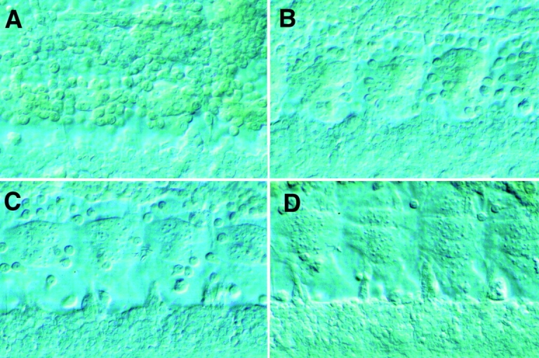
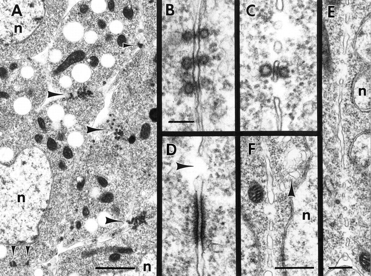
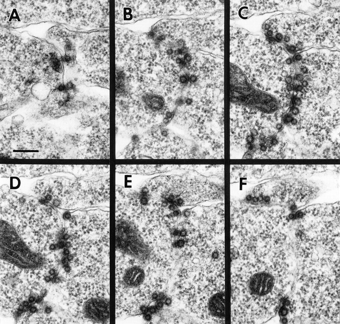
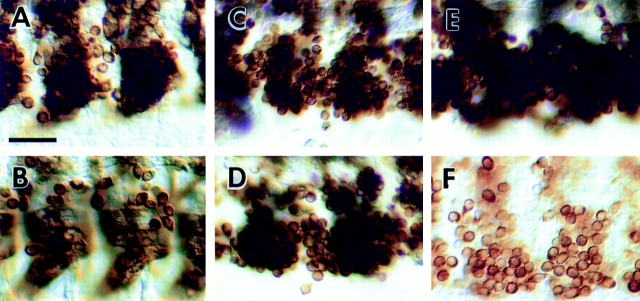
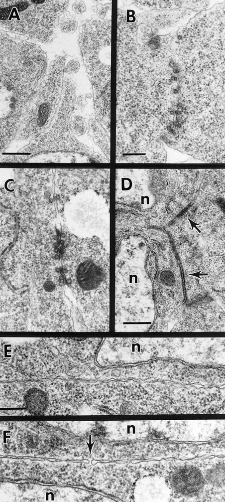
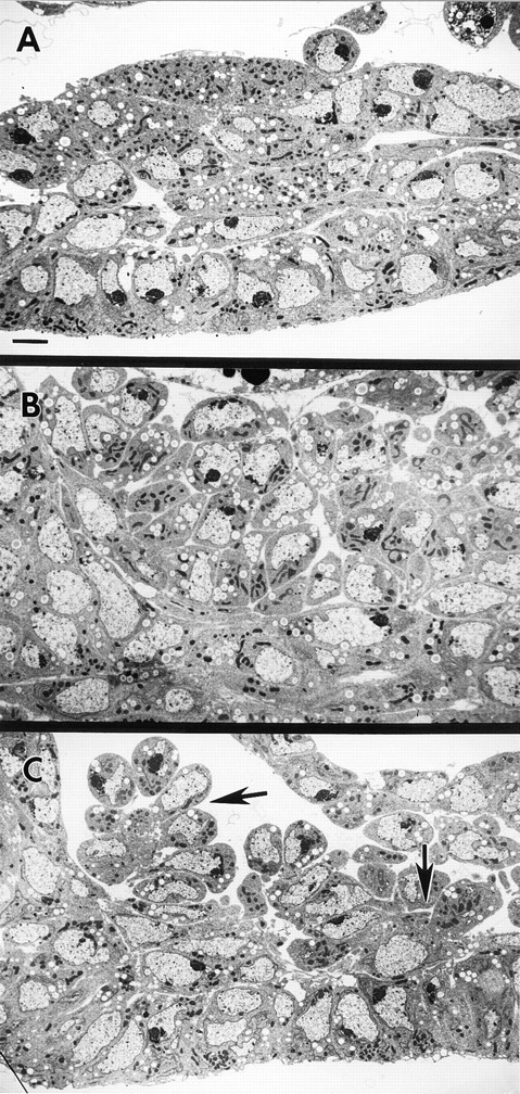
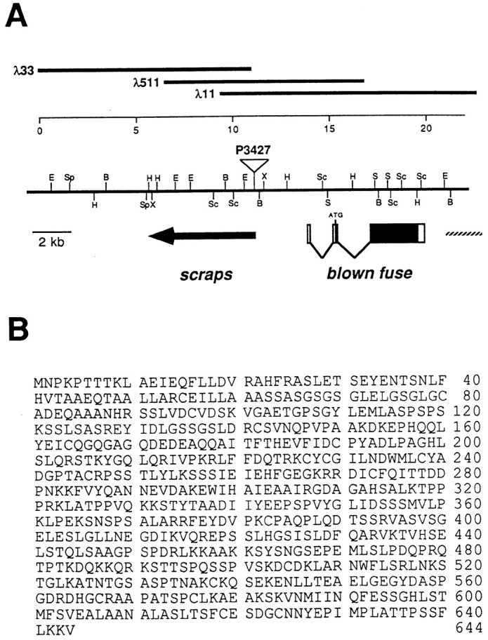
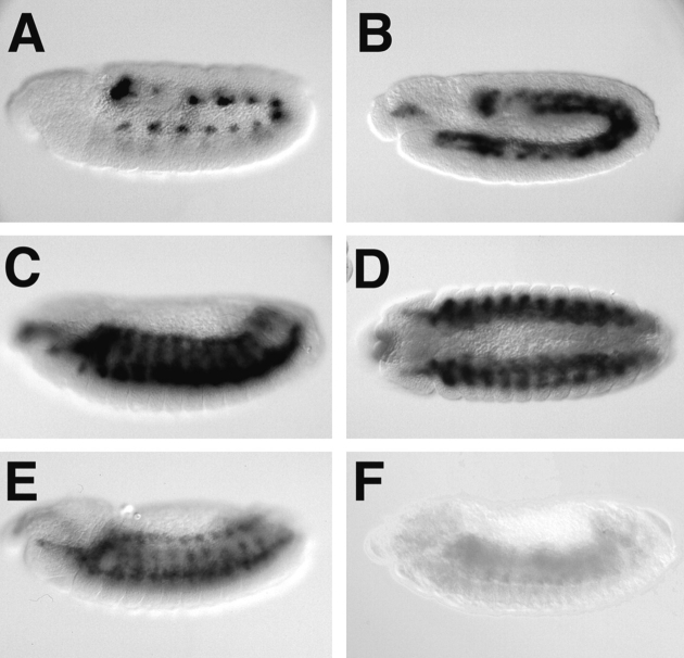
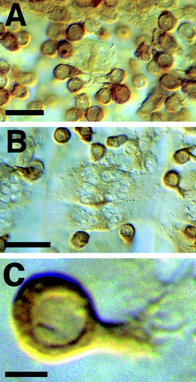
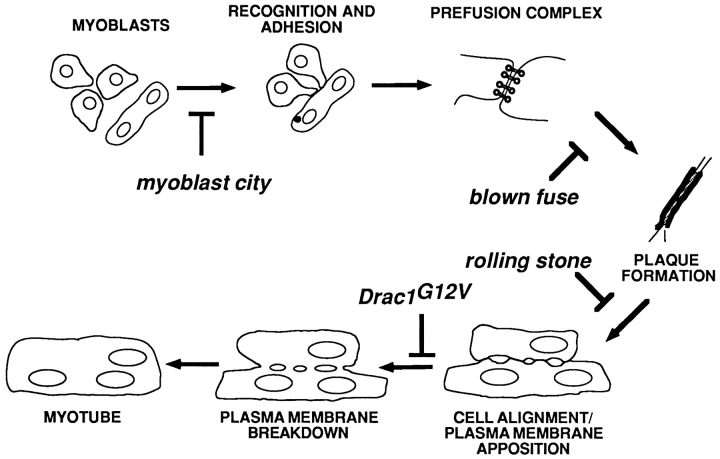
The events of myoblast fusion in Drosophila are dissected here by combining genetic analysis with light and electron microscopy. We describe a new and essential intermediate step in the process, the formation of a prefusion complex consisting of "paired vesicles." These pairs of vesicles from different cells align with each other across apposed plasma membranes. This prefusion complex resolves into dense membrane plaques between apposed cells; these cells then establish cytoplasmic continuity by fusion of small areas of plasma membrane followed by vesiculation of apposed membranes. Different steps in this process are specifically blocked by mutations in four genes required for myoblast fusion. One of these genes, blown fuse, encodes a novel cytoplasmic protein expressed in unfused myoblasts that is essential for progression beyond the prefusion complex stage.


Figures

Figure 1
Figure 1
Myoblast fusion in the developing Drosophila embryo. Light level micrographs of myoblast fusion in the ventral muscle region of wild-type Drosophila embryos. Developing muscles are imaged by Nomarski optics, and the plane of focus is close to the epidermis. (A) Wild-type early stage 13 embryo. Small early myotubes are present, with many unfused myoblasts attached to the surface of the myotubes. (B) Wild-type stage 14 embryo. (C) Wild-type stage 15 embryo. Myotubes are substantially larger, with few unfused myoblasts remaining. (D) Wild-type stage 16 embryo.
Figure 2
Figure 2
Ultrastructure of intermediate steps in myoblast fusion. Electron micrographs of wild-type myoblast fusion in early stage 13 embryos. All stages of the fusion process occur simultaneously in various parts of the developing musculature. (A) Myoblasts in early stage of fusion. Note prefusion complexes at points of cell–cell contact (arrowheads); n indicates myoblast nuclei. (B) Three sets of paired vesicles. Note electron-dense material in the extracellular space between pairs of vesicles. (C) Paired vesicles oriented across a vesiculating pair of plasma membranes. (D) An electron-dense plaque near a region of actively fusing membrane; note fusion pore (arrow). (E) Fusion pores in a vesiculating plasma membrane. The cytoplasm within and beneath the pore is free of staining material such as ribosomes. (F) Later stage vesiculating plasma membrane. The membrane sacs have increased in width and a group of irregular clear vesicles is present (arrowhead). Bars: (A) 1 μm; (B–D) 100 nm; (E) 250 μm; (F) 500 μm.
Figure 3
Figure 3
The prefusion complex contains paired vesicles. Serial section electron micrographs through a prefusion complex in a wild-type stage 13 embryo. This complex contains about 45 pairs of vesicles distributed among three cells. Bar: (A) 200 nm.
Figure 4
Figure 4
Mutations in genes that are essential for myoblast fusion. Light level micrographs of myoblast fusion in the ventral muscle region of wild-type (A–B) and mutant (C–F) Drosophila embryos. Myoblasts are stained with anti-myosin monoclonal antibody FMM5 (Kiehart and Feghali, 1986). The plane of focus is more superficial (closer to the gut) than in Fig. 1 to discern individual unfused myoblasts. (A) Wild-type stage 13 embryo. Fusion has begun and the early ventral myotubes are beginning to extend towards their attachment sites. (B) Wild-type stage 14 embryo. Myotubes have attached to the epidermis and unfused myoblasts are present on the superficial surface of the myotubes. (C) mbcC1 stage 14 embryo. Compare to B; little or no fusion has occurred. (D) blow2 stage 14 embryo. The myoblasts are more tightly clustered than in mbc mutants. (E) rost15 stage 14 embryo. The morphology of the unfused myoblast clusters is different from other mutants due to the alignment of myoblasts close to the epidermis. (F) Drac1G12V:GAL4-24B stage 14 embryo. Most of the unfused myoblasts have been removed by macrophages. Bar, 25 μm.
Figure 6
Figure 6
Different mutants block specific steps in myoblast fusion. (A) Representative cell–cell contacts between myoblasts in an early stage 13 mbcC1 mutant embryo. Prefusion complexes are absent. (B) Prefusion complex in a stage 13 blow2 mutant embryo. The complexes in this mutant are indistinguishable from those in wild-type embryos. (C) Prefusion complex in a stage 13 rost4 mutant embryo. The prefusion complexes in this mutant are also indistinguishable from wild type. (D) Membrane plaques (arrows) between three myoblasts in a rost15 embryo. n indicates myoblast nuclei. (E) Close apposition of plasma membranes in a stage 14 rost15 embryo. (F) Abortive plasma membrane fusion in a Drac1G12V/24B embryo. A single fusion pore is visible (arrow). At certain places the apposed plasma membranes are so close, they are indistinguishable from a single membrane. Bars: (A) 500 nm; (B and C) 250 nm; (D) 500 nm; (E and F) 250 nm.
Figure 5
Figure 5
mbc is required for recognition and/or attachment of pioneer cells by myoblasts. Electron micrographs of ventral muscle region in stage 14 homozygous embryos. (A) Wild-type embryo. The ventral nerve cord is to the left side of the frame. (B) mbcC1 embryo. The unfused myoblasts are oriented in an apparently random manner, indicating that recognition and/or attachment of myoblasts to pioneer cells is disrupted in this mutant. (C) blow2 embryo. Note groups of myoblasts attached to single pioneer cells (arrows). Bar, 2 μm.
Figure 7
Figure 7
Genomic organization and sequence of the blown fuse gene. (A) Genomic map of the region in 43E containing scraps and blown fuse. At top, numbers represent scale in kilobases. Thick lines indicate representative λ phage clones isolated during the chromosomal walk. Single letter abbreviations indicate restriction sites: B, BamHI; E, EcoRI; H, HindIII; S, SalI; Sc, SacI; Sp, SpeI; X, XbaI. The insertion site of scrapsP3427 is indicated by an open triangle. Beneath the chromosomal map, the location of the scraps gene is indicated by a solid arrow. The blown fuse gene is represented by boxes, with open boxes indicating noncoding regions and solid boxes indicating the coding region. Part of an unidentified third gene is indicated by a hatched line. (B) Amino acid sequence of the Blown Fuse protein.
Figure 8
Figure 8
Expression of blow mRNA. Expression pattern of blow mRNA. (A) In situ hybridization of the blow cDNA to a whole mount stage 10 embryo. The mRNA is expressed in 12 cell clusters in the developing mesoderm. (B) In situ hybridization to a stage 12 embryo. (C) In situ hybridization to a stage 13 embryo. The mRNA is expressed at high levels in myoblasts, and is not expressed in other cells. (D) In situ hybridization to a stage 13 embryo, ventral view. (E) In situ hybridization to a stage 14 embryo. Expression level is lower than in previous stages. (F) In situ hybridization to a homozygous blow2 stage 13 embryo. No mRNA is detectable.
Figure 9
Figure 9
Localization of Blow protein. Subcellular localization of Blow protein. (A and B) Anti-Blow staining in a dissected stage 13 wild-type embryo. Little protein is present in myotubes relative to the level in unfused myoblasts. (C) Anti-Blow staining in a wild-type myoblast. The protein is distributed evenly throughout the cytoplasm of the myoblasts and is excluded from the nucleus. Note the large single pseudopodium. Bars: (A and B) 10 μm; (C) 2 μm.
Figure 10
Figure 10
Model of intermediate steps in myoblast fusion. Proposed schematic of the steps of myoblast fusion at the ultrastructural level, indicating action points of each mutant.

References

    1. Abmayr SM, Erickson MS, Bour BA. Embryonic development of the larval body wall musculature of Drosophila melanogaster. . Trends Genet. 1995;11:153–159. - PubMed
    1. Altschul SF, Gish W, Miller W, Myers EW, Lipman DJ. Basic local alignment search tool. J Mol Biol. 1990;215:403–410. - PubMed
    1. Ball EE, Goodman CS. Muscle development in the grasshopper embryo: II. Syncytial origin of the extensor tibiae muscle pioneers. Dev Biol. 1985a;111:399–416. - PubMed
    1. Ball EE, Goodman CS. Muscle development in the grasshopper embryo: III. Sequential origin of the flexor tibiae muscle pioneers. Dev Biol. 1985b;111:417–424. - PubMed
    1. Ball EE, Ho RK, Goodman CS. Muscle development in the grasshopper embryo. I. Muscles, nerves and apodemes in the metathoracic leg. Dev Biol. 1985c;111:383–398. - PubMed

Publication types