. 1997 Mar 24;136(6):1263-70.
doi: 10.1083/jcb.136.6.1263.

beta-Actin messenger RNA localization and protein synthesis augment cell motility

Affiliations

beta-Actin messenger RNA localization and protein synthesis augment cell motility

E H Kislauskis et al. J Cell Biol. .

Erratum in

  • J Cell Biol 1997 Jun 30;137(7):1683

Abstract

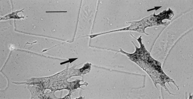
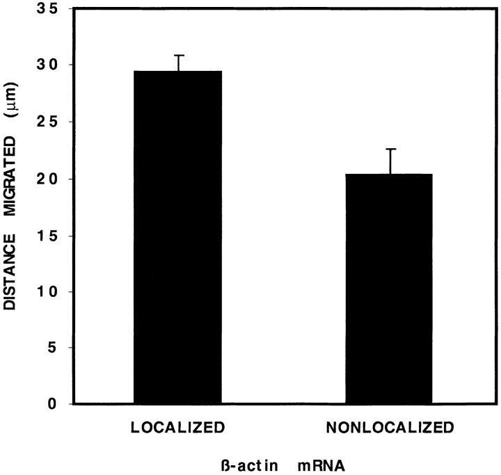
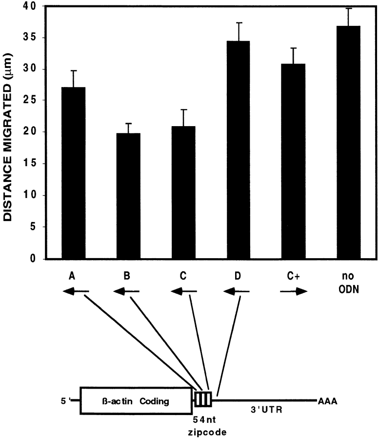
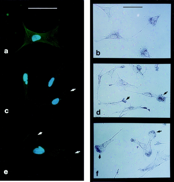
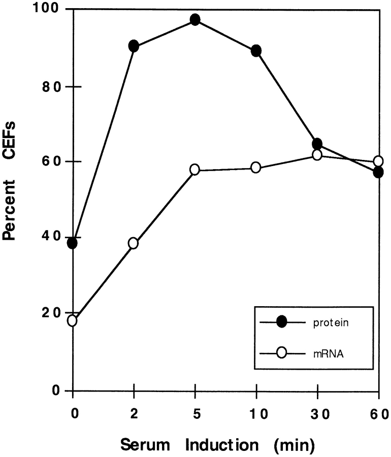
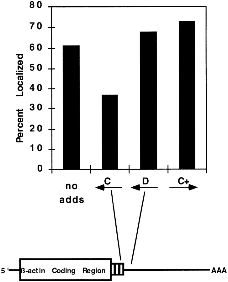
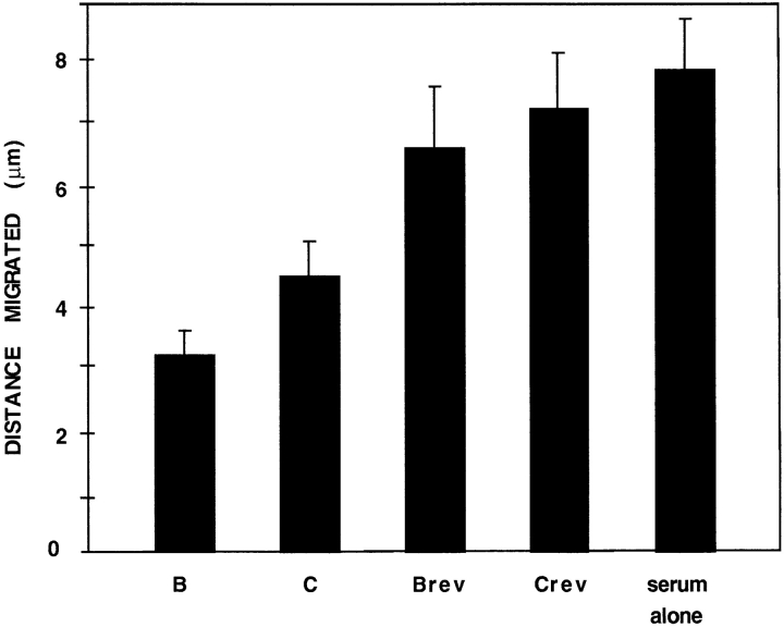
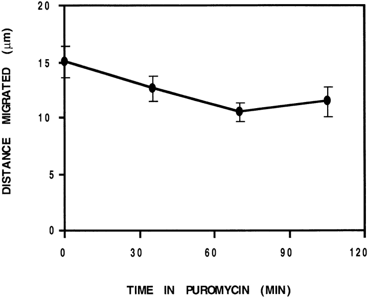
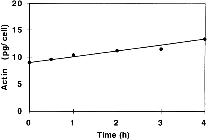
In chicken embryo fibroblasts (CEFs), beta-actin mRNA localizes near an actin-rich region of cytoplasm specialized for motility, the lamellipodia. This localization is mediated by isoform-specific 3'-untranslated sequences (zipcodes) and can be inhibited by antizipcode oligodeoxynucleotides (ODNs) (Kislauskis, E.H., X.-C. Zhu, and R.H. Singer. 1994. J. Cell Biol. 127: 441-451). This inhibition of beta-actin mRNA localization resulted in the disruption of fibroblast polarity and, presumably, cell motility. To investigate the role of beta-actin mRNA in motility, we correlated time-lapse images of moving CEFs with the distribution of beta-actin mRNA in these cells. CEFs with localized beta-actin mRNA moved significantly further over the same time period than did CEFs with nonlocalized mRNA. Antizipcode ODN treatment reduced this cell translocation while control ODN treatments showed no effect. The temporal relationship of beta-actin mRNA localization to cell translocation was investigated using serum addition to serum-deprived cultures. beta-actin mRNA was not localized in serum-deprived cells but became localized within minutes after serum addition (Latham, V.M., E.H. Kislauskis, R.H. Singer, and A.F. Ross. 1994. J. Cell Biol. 126:1211-1219). Cell translocation increased over the next 90 min, and actin synthesis likewise increased. Puromycin reduced this cell translocation and blocked this induction in cytosolic actin content. The serum induction of cell movement was also inhibited by antizipcode ODNs. These observations support the hypothesis that beta-actin mRNA localization and consequent protein synthesis augment cell motility.


Figures

Figure 1
Figure 1
Direction of cell motility correlates with β-actin distribution. CEFs cultured on gelatin-coated, gridded coverslips were video recorded twice, 45 min apart, and then fixed and processed to detect β-actin mRNA by in situ hybridization. Alkaline phosphatase activity (dark intracellular staining) corresponded to the distribution of endogenous β-actin mRNA at the time of fixation. Signal is well-localized in the lamellae of two CEFs (in the direction of motility, arrows) and not in a third. Grid square is 175 μm. Bar, 30 μm.
Figure 2
Figure 2
Speed correlates with localization of β-actin mRNA. CEF speed was calculated for 171 CEFs from the magnitude of the distance each CEF migrated (translocated) during the 45–60min period. Nearly all CEFs (93%) moved during that period. β-actin mRNA was localized (predominantly peripheral) in 68% of CEFs. CEFs with localized β-actin mRNA migrated 1.7-fold faster than nonlocalized. Error bars indicate the standard error of the mean for each data point.
Figure 3
Figure 3
Antizipcode oligodeoxynucleotides inhibit cell motility. CEFs were treated for 12 h with various phosphorothioate ODNs corresponding to antisense (A–D), sense (C+), or no ODNs, as depicted schematically. A significant reduction in translocation over a 60-min period was observed with ODNs complementary to the middle and distal third of the 54-nucleotide zipcode, only (B or C). Error bars indicate the standard error of the mean for each data point.
Figure 4
Figure 4
Serum-induced relocalization of β-actin protein and its mRNA. CEFs cultured on gelatin coverslips were serum deprived for 24 h and induced with 10% FBS and fixed after 2 and 5 min. Representative examples of the distribution of β-actin–specific FITCimmunocytochemistry (a, c, and e) and β-actin mRNA by in situ hybridization (b, d, and f) are shown. a and b, 24 h starved control; c and d, 2 min induced; and e and f, 5 min induced. Arrowheads show lamellipodia staining and membrane protrusion. Bar, 20 μm.
Figure 5
Figure 5
The course of serum-induced relocalization of β-actin protein and its mRNA. CEFs were induced with 10% FBS after a 24 h serum starvation (Fig. 4). Coverslips with CEFs were removed 0, 2, 5, 10, 30, and 60 min after induction. The percent of CEFs is represented with localized mRNA (open circles) or with lamellipodia staining by β-actin immunocytochemistry (closed circles). The experimental mean was calculated from three separate experiments for each point except 60 min (single experiment).
Figure 6
Figure 6
Serum-induced relocalization of β-actin mRNA is mediated through the β-actin zipcode. CEFs were serum deprived for 24 h and induced for 30 min with 10% FBS containing the indicated ODNs or no ODNs. β-Actin mRNA localization in the population was evaluated by in situ hybridization as described in Materials and Methods. The effects of ODNs on the percentage of CEF with localized β-actin mRNA signal is shown. The serumstarved level of localization was 23%.
Figure 7
Figure 7
Serum induction of motility is inhibited by antisense ODNs. CEFs were serum deprived for 24 h and induced with 10% FBS containing the indicated ODN for 90 min as indicated. Motility (translocation) was measured over a 30-min interval after treatment. Similar to the results in Fig. 3, antizipcode ODN B significantly inhibited translocation, while control ODN Brev did not.
Figure 8
Figure 8
Inhibition of protein synthesis suppresses motility. Analysis of translocation distance for a steady-state culture of CEFs over consecutive 30-min periods after treatment with puromycin (200 μg/ml). Control cells were evaluated before treatment (time 0). Translocation was suppressed by 30% after 70 min in puromycin.
Figure 9
Figure 9
Cytoplasmic actin content after serum induction. Line plot of the increase in actin content in serum-starved CEFs before and after the addition of serum. Coomassie-stained gels were used for quantitation.

References

    1. Albrecht-Buhler G. Autonomous movements of cytoplasmic fragments. Proc Natl Acad Sci USA. 1980;77:6639–6643. - PMC - PubMed
    1. Bershadsky AD, Gluck U, Denisenko ON, Sklyarova TV, Spector I, Ben-Ze'ev A. The state of actin assembly regulates actin and vinculin expression by a feedback loop. J Cell Sci. 1995;108:1183–1193. - PubMed
    1. Condeelis J. Life at the leading edge: the formation of cell protrusions. Annu Rev Cell Biol. 1993;9:411–444. - PubMed
    1. Darnell, J.E., H. Lodish, D. Baltimore, A. Berk, S.L. Zipursky, and P. Matsudaira. 1995. Molecular Cell Biology. W. H. Freeman, editor. Scientific American Books, Inc., New York. 129.
    1. Farmer SR, Wan KM, Ben-Ze'ev A, Penman S. Regulation of actin mRNA levels and translation responds to changes in cell configuration. Mol Cell Biol. 1983;3:182–189. - PMC - PubMed

Publication types

MeSH terms