. 1997 Mar 24;136(6):1323-32.
doi: 10.1083/jcb.136.6.1323.

Xenopus actin depolymerizing factor/cofilin (XAC) is responsible for the turnover of actin filaments in Listeria monocytogenes tails

Affiliations

Xenopus actin depolymerizing factor/cofilin (XAC) is responsible for the turnover of actin filaments in Listeria monocytogenes tails

J Rosenblatt et al. J Cell Biol. .

Abstract

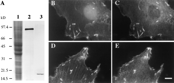
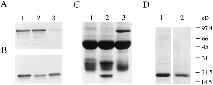
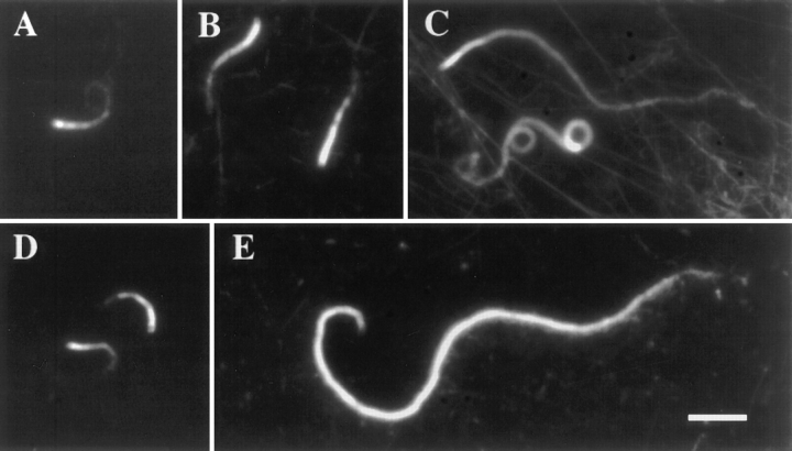
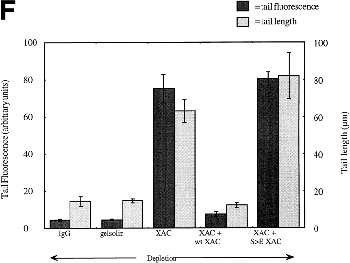
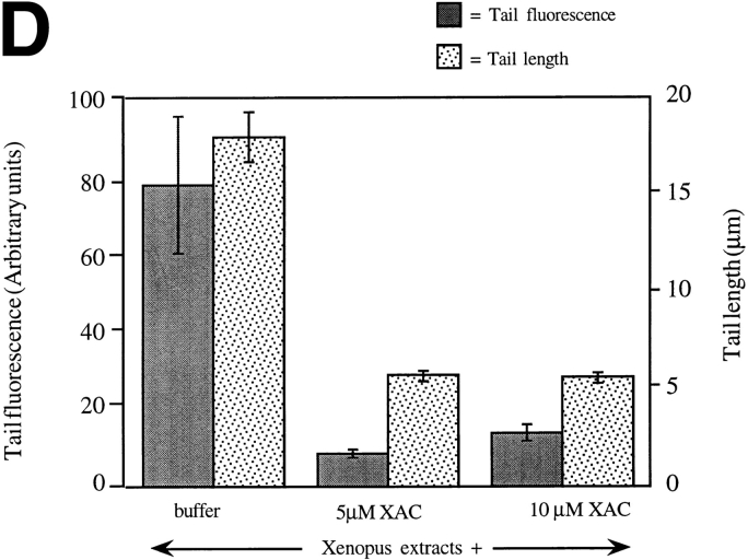
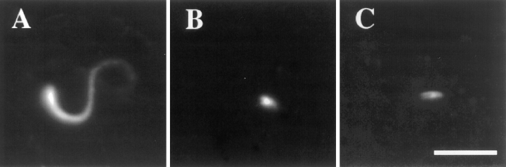
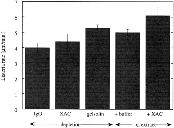
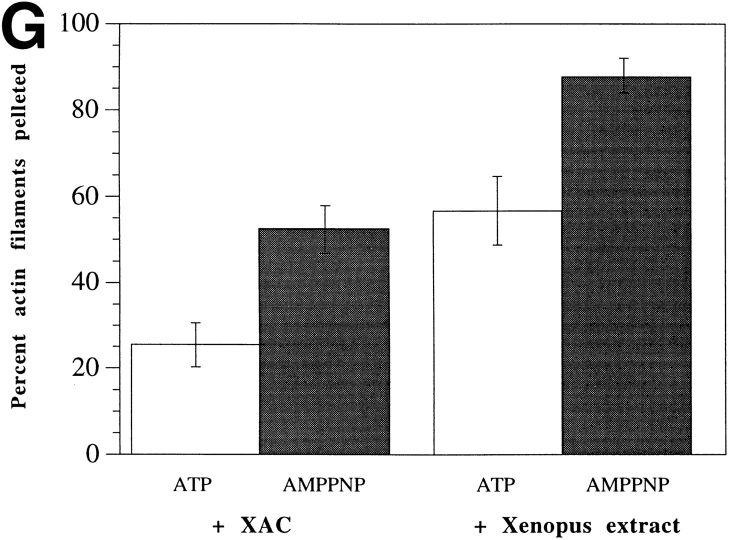
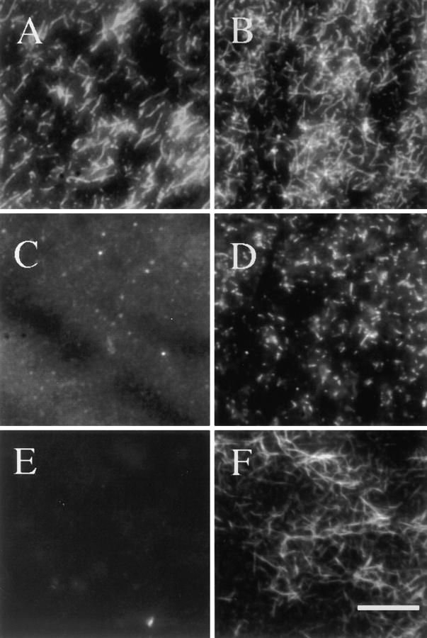
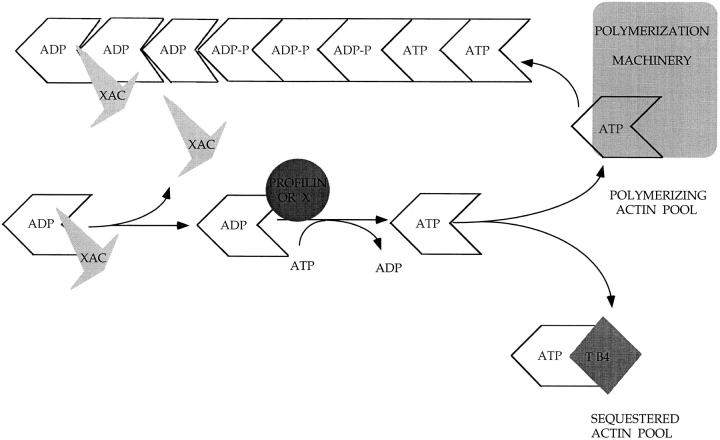
In contrast to the slow rate of depolymerization of pure actin in vitro, populations of actin filaments in vivo turn over rapidly. Therefore, the rate of actin depolymerization must be accelerated by one or more factors in the cell. Since the actin dynamics in Listeria monocytogenes tails bear many similarities to those in the lamellipodia of moving cells, we have used Listeria as a model system to isolate factors required for regulating the rapid actin filament turnover involved in cell migration. Using a cell-free Xenopus egg extract system to reproduce the Listeria movement seen in a cell, we depleted candidate depolymerizing proteins and analyzed the effect that their removal had on the morphology of Listeria tails. Immunodepletion of Xenopus actin depolymerizing factor (ADF)/cofilin (XAC) from Xenopus egg extracts resulted in Listeria tails that were approximately five times longer than the tails from undepleted extracts. Depletion of XAC did not affect the tail assembly rate, suggesting that the increased tail length was caused by an inhibition of actin filament depolymerization. Immunodepletion of Xenopus gelsolin had no effect on either tail length or assembly rate. Addition of recombinant wild-type XAC or chick ADF protein to XAC-depleted extracts restored the tail length to that of control extracts, while addition of mutant ADF S3E that mimics the phosphorylated, inactive form of ADF did not reduce the tail length. Addition of excess wild-type XAC to Xenopus egg extracts reduced the length of Listeria tails to a limited extent. These observations show that XAC but not gelsolin is essential for depolymerizing actin filaments that rapidly turn over in Xenopus extracts. We also show that while the depolymerizing activities of XAC and Xenopus extract are effective at depolymerizing normal filaments containing ADP, they are unable to completely depolymerize actin filaments containing AMPPNP, a slowly hydrolyzible ATP analog. This observation suggests that the substrate for XAC is the ADP-bound subunit of actin and that the lifetime of a filament is controlled by its nucleotide content.


Figures

Figure 1
Figure 1
Localization of XAC and gelsolin in Listeria-infected XL 177 cells. (A) Western blot analysis. Lane 1, Coomassie blue–stained SDS-PAGE gel of total XL 177 cell lysate; lane 2, XL 177 lysate probed with anti-Xenopus gelsolin antibody; lane 3, XL 177 lysate probed with anti-XAC antibody. Immunostaining of Listeria-infected XL 177 cells with gelsolin antibody (C) or XAC antibody (E). F-actin in B and D is visualized with fluorescein-phalloidin. Both gelsolin and XAC colocalize with F-actin in Listeria tails. Bar, 10 μm.
Figure 2
Figure 2
Gels of immunodepletion of XAC and gelsolin from Xenopus laevis egg extracts and purified XAC and ADF. (A and B) Immunoblots of immunodepleted extracts using antibodies to gelsolin (A) and XAC (B). For both A and B, lane 1 is the IgG-depleted control, lane 2 is the XAC-depleted extract, and lane 3 is the gelsolin-depleted extract. Quantitation of the depletion was performed by densitometry of the bands in A and B compared to a dilution series of pure extract. (C) Coomassie blue–stained SDS-PAGE gel of immunoprecipitated complexes with XAC and gelsolin antibodies. Lane 1, the heavy and light chain of random rabbit IgG alone; lane 2, XAC (19 kD) and another band at approximately 28 kD over the IgG heavy and light chain bands; lane 3, gelsolin (∼93 kD) over the IgG bands. (D) Coomassie blue–stained SDS-PAGE shows the purity of the recombinant XAC and chicken ADF mutant that were added back to the XAC immunodepletions. Lane 1, wild-type XAC; lane 2, S3E ADF. Apparent molecular mass markers for all gels are indicated on the right.
Figure 3
Figure 3
Listeria tail formed in XAC- or gelsolin-depleted Xenopus egg extracts. Actin tails are visualized by mixing rhodaminelabeled actin to Listeria and the following extracts: (A) IgG- depleted extracts, (B) gelsolin-depleted extracts, (C) XAC- depleted extracts, (D) XAC-depleted extracts plus 2.7 μM pure wild-type XAC, and (E) XAC-depleted extracts plus 2.7 μM pure S3E ADF. (F) Bar graph quantitating the amount of tail fluorescence (left side, ▒⃞ ▒⃞ ) and tail length (right side, ░⃞ ) using Winview software. The number of tails analyzed for IgG depletion was 33, for gelsolin depletion, 29, for XAC depletion, 43, for wildtype XAC addback, 25, and for S3E ADF addback, 11, over seven experiments using two separate extracts preps. Error bars represent standard deviation of the mean. Bar, 10 μm.
Figure 3
Figure 3
Listeria tail formed in XAC- or gelsolin-depleted Xenopus egg extracts. Actin tails are visualized by mixing rhodaminelabeled actin to Listeria and the following extracts: (A) IgG- depleted extracts, (B) gelsolin-depleted extracts, (C) XAC- depleted extracts, (D) XAC-depleted extracts plus 2.7 μM pure wild-type XAC, and (E) XAC-depleted extracts plus 2.7 μM pure S3E ADF. (F) Bar graph quantitating the amount of tail fluorescence (left side, ▒⃞ ▒⃞ ) and tail length (right side, ░⃞ ) using Winview software. The number of tails analyzed for IgG depletion was 33, for gelsolin depletion, 29, for XAC depletion, 43, for wildtype XAC addback, 25, and for S3E ADF addback, 11, over seven experiments using two separate extracts preps. Error bars represent standard deviation of the mean. Bar, 10 μm.
Figure 4
Figure 4
Listeria tails resulting from addition of excess XAC to Xenopus egg extracts. (A) Actin tail resulting from adding extract buffer to Xenopus egg extracts. (B) Actin tails resulting from adding wild-type XAC to extracts to 7.1 μM. (C) Actin tails resulting from adding wild-type XAC to 12.1 μM. (D) Quantitative analysis of the amount of tail fluorescence (left side, ▒⃞ ░⃞ ) and tail length (right side, ░⃞ ) using Winview software. The fluorescence units differ from those in Fig. 3 because the images were analyzed at different magnifications. The number of tails analyzed for buffer alone addition was 15, for XAC addition to 7.1 μM, 29, and for XAC addition to 12.1 μM, 33, over three experiments using two separate extract preps. Error bars represent standard deviation of the mean. Bar, 10 μm.
Figure 4
Figure 4
Listeria tails resulting from addition of excess XAC to Xenopus egg extracts. (A) Actin tail resulting from adding extract buffer to Xenopus egg extracts. (B) Actin tails resulting from adding wild-type XAC to extracts to 7.1 μM. (C) Actin tails resulting from adding wild-type XAC to 12.1 μM. (D) Quantitative analysis of the amount of tail fluorescence (left side, ▒⃞ ░⃞ ) and tail length (right side, ░⃞ ) using Winview software. The fluorescence units differ from those in Fig. 3 because the images were analyzed at different magnifications. The number of tails analyzed for buffer alone addition was 15, for XAC addition to 7.1 μM, 29, and for XAC addition to 12.1 μM, 33, over three experiments using two separate extract preps. Error bars represent standard deviation of the mean. Bar, 10 μm.
Figure 5
Figure 5
Bar graph representing analysis of Listeria movement rate during various extract treatments. The rates were measured using Image 1 software or Winview software. The number of tails measured for each treatment were: IgG depletion, 25; XAC depletion, 24; gelsolin depletion, 34; buffer addition, 27, and XAC addition (to 5.0 μM), 24, over four experiments using two separate extract preps. Error bars represent standard deviation of the mean.
Figure 6
Figure 6
The resistance of AMPPNP–containing actin filaments to the depolymerizing activities of XAC and Xenopus egg extracts. Approximately 0.5 μM rhodamine-labeled actin polymerized with either ATP (A, C, and E) or AMPPNP (B, D, and F). Actin filaments mixed 1:1 with F-buffer (A and B), with 5.3 μM XAC (C and D), and with Xenopus extract (E and F). In both cases (C–F), XAC is in excess of rhodamine F-actin by approximately fivefold. (G) Quantitation of the amount of rhodamine- labeled ATP (□) versus AMPPNP ( ▒⃞ ░⃞ ) F-actin pelleted in the presence of XAC or Xenopus egg extracts. Percent actin filaments remaining was calculated as the fluorescence of the rhodamine F-actin pelleted in XAC or extracts/the fluorescence of rhodamine F-actin pelleted in F-buffer. The bars represent an average of four experiments and the error bars represent standard deviation of the mean. Bar, 10 μm.
Figure 6
Figure 6
The resistance of AMPPNP–containing actin filaments to the depolymerizing activities of XAC and Xenopus egg extracts. Approximately 0.5 μM rhodamine-labeled actin polymerized with either ATP (A, C, and E) or AMPPNP (B, D, and F). Actin filaments mixed 1:1 with F-buffer (A and B), with 5.3 μM XAC (C and D), and with Xenopus extract (E and F). In both cases (C–F), XAC is in excess of rhodamine F-actin by approximately fivefold. (G) Quantitation of the amount of rhodamine- labeled ATP (□) versus AMPPNP ( ▒⃞ ░⃞ ) F-actin pelleted in the presence of XAC or Xenopus egg extracts. Percent actin filaments remaining was calculated as the fluorescence of the rhodamine F-actin pelleted in XAC or extracts/the fluorescence of rhodamine F-actin pelleted in F-buffer. The bars represent an average of four experiments and the error bars represent standard deviation of the mean. Bar, 10 μm.
Figure 7
Figure 7
Model for recycling of actin by ADF/cofilin proteins. At areas of high filament turnover in the cell, ATP-bound actin is induced to polymerize by a complex of proteins. The ATP within the filament is hydrolyzed and the terminal phosphate is slowly released. The resulting ADP-containing subunits have weaker interactions with each other than those containing ATP. These subunits are now available for depolymerization by the ADF/cofilin family of proteins (XAC). Once XAC depolymerizes an actin subunit(s), it dissociates from actin. The released actin must exchange its ADP for ATP and the nucleotide exchange is probably catalyzed by profilin. The ATP-bound actin is then either repolymerized or sequestered for later use.

Comment in

References

    1. Abe H, Obinata T, Minamide LS, Bamburg JR. Xenopus laevisactin-depolymerizing factor/cofilin: a phosphorylation-regulated protein essential for development. J Cell Biol. 1996;132:871–885. - PMC - PubMed
    1. Agnew BJ, Minamide LS, Bamburg JR. Reactivation of phosphorylated actin depolymerizing factor and identification of the regulatory site. J Biol Chem. 1995;270:17582–17587. - PubMed
    1. Ampe C, Vandekerckhove J. The F-actin capping proteins of Physarum polycephalum: cap42(a) is very similar, if not identical, to fragmin and is structurally and functionally very homologous to gelsolin; cap42(b) is Physarum actin. EMBO (Eur Mol Biol Organ) J. 1987;6:4149–4157. - PMC - PubMed
    1. André E, Lottspeich F, Schleicher M, Noegel A. Severin, gelsolin, and villin share a homologous sequence in regions presumed to contain F-actin severing domains. J Biol Chem. 1988;263:722–727. - PubMed
    1. André E, Brink M, Gerisch G, Isenberg G, Noegel A, Schleicher M, Segall JE, Wallraff E. A Dictyosteliummutant deficient in severin, an F-actin fragmenting protein, shows normal motility and chemotaxis. J Cell Biol. 1989;108:985–995. - PMC - PubMed

Publication types

MeSH terms