. 1997 Mar 24;136(6):1363-73.
doi: 10.1083/jcb.136.6.1363.

Matrix metalloproteinases MMP2 and MMP9 are produced in early stages of kidney morphogenesis but only MMP9 is required for renal organogenesis in vitro

Affiliations

Matrix metalloproteinases MMP2 and MMP9 are produced in early stages of kidney morphogenesis but only MMP9 is required for renal organogenesis in vitro

B Lelongt et al. J Cell Biol. .

Abstract

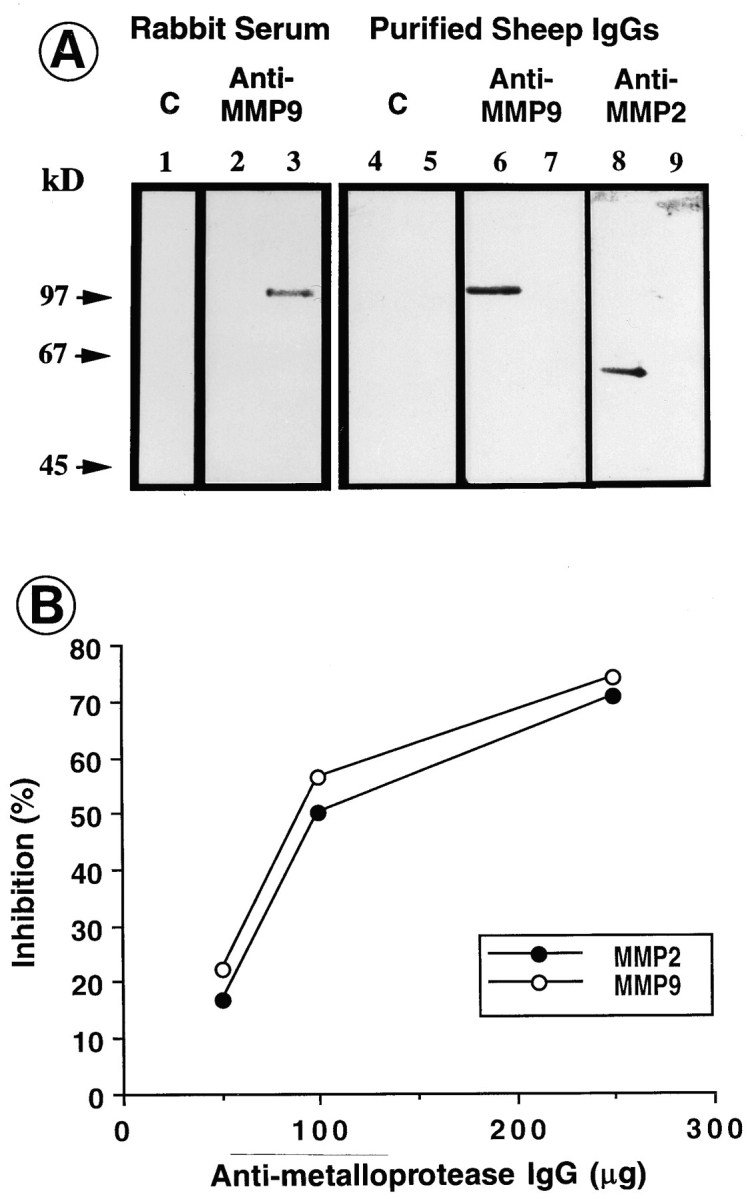
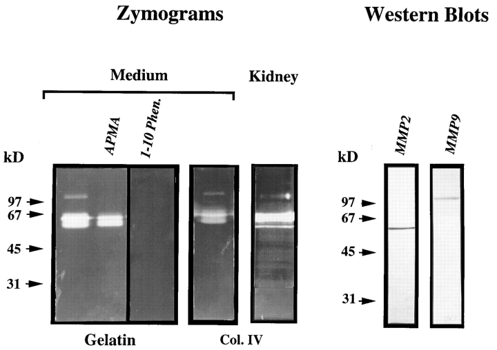
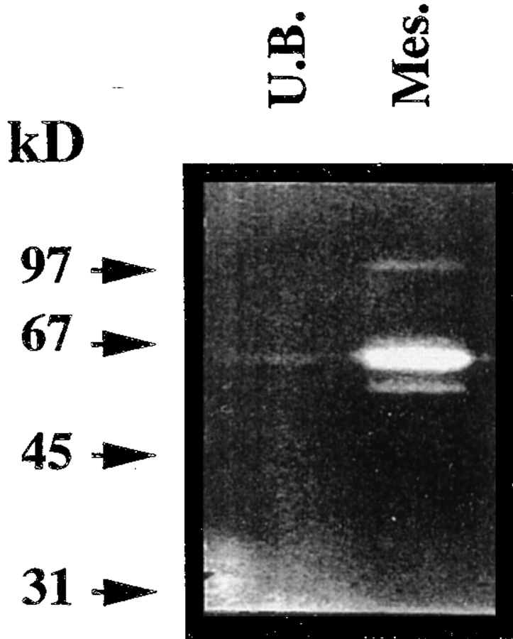
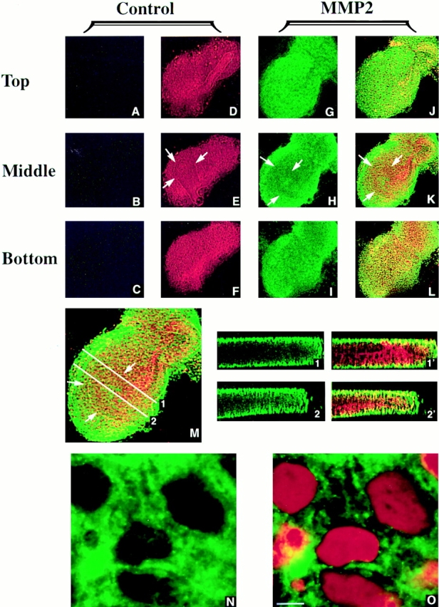
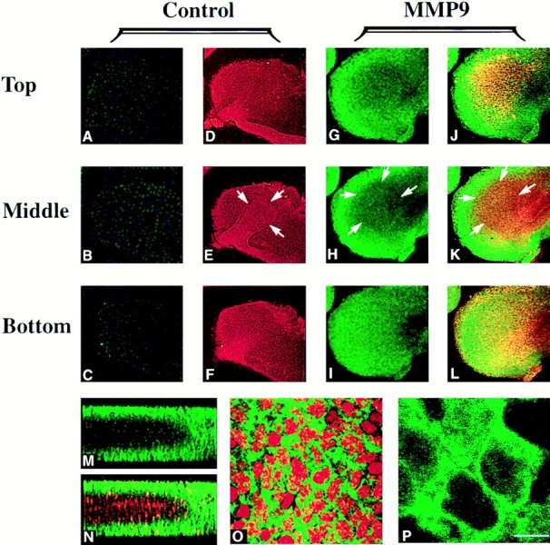
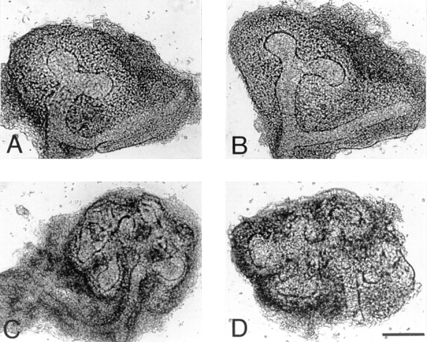
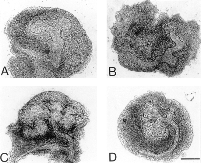
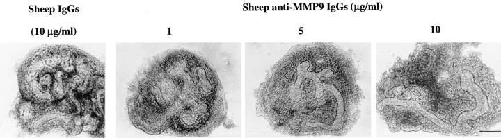
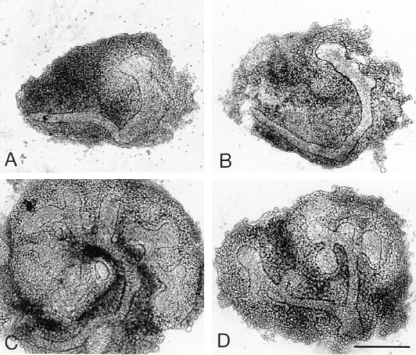
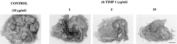
We analyzed matrix metalloproteinase (MMP) production by 11-d embryonic mouse kidneys and the effects of these enzymes on subsequent renal organogenesis. In vivo, immunolocalization of metalloproteinases by laser scanning confocal microscopy and zymograms of kidney lysates showed that the mesenchyme of embryonic kidneys synthesized both MMP9 and MMP2 enzymes. In vitro, embryonic kidneys also secreted both enzymes when cultured in a medium devoid of hormone, growth factor, and serum for 24 h during which T-shaped branching of the ureter bud appeared. We then evaluated the role of MMP2 and MMP9 in kidney morphogenesis by adding anti-MMP2 or anti-MMP9 IgGs to the culture medium of 11-d kidneys for 24 or 72 h. Although it inhibited activity of the mouse enzyme, anti-MMP2 IgGs had no effect on kidney morphogenesis. In contrast, anti-MMP9 IgGs with enzyme-blocking activity impaired renal morphogenesis, in a concentration-dependent manner, by inhibiting T-shaped branching and further divisions of the ureter bud. This effect was irreversible, still observed after inductive events and reproduced by exogenous tissue inhibitor of metalloproteinase 1 (TIMP1), the natural inhibitor of MMP9. These data provide the first demonstration of MMP9 and MMP2 production in vivo by 11-d embryonic kidneys and further show that MMP9 is required in vitro for branching morphogenesis of the ureter bud.


Figures

Figure 1
Figure 1
Antibody characterization. (A) Western blots performed with MMP9 (lanes 1, 3, 4, 6, and 9) and MMP2 (lanes 2, 5, 7, and 8) isolated from mouse macrophage and NIH 3T3 cell lines, respectively. Rabbit anti–MMP9 antibody (lanes 2 and 3) and sheep anti–MMP9 IgGs (lanes 6 and 7) recognized mouse macrophage MMP9 (lanes 3 and 6) but not mouse fibroblast MMP2 (lanes 2 and 7). Conversely, sheep anti–MMP2 IgGs (lanes 8 and 9) recognized mouse fibroblast MMP2 (lane 8) but not mouse macrophage MMP9 (lane 9). Neither rabbit preimmune serum (lane 1) nor sheep preimmune IgGs (lanes 4 and 5) reacted with mouse MMP9 (lanes 1 and 4) or MMP2 (lane 5). (B) The inhibitory effect of antibodies on enzymatic activity was estimated by incubating an aliquot (0.6 U) of MMP2 or MMP9 with heat-denaturated 14C-type I collagen in the presence or absence of increasing amounts of sheep anti–MMP2 or anti–MMP9 IgGs or of sheep preimmune IgGs. Results are expressed as the percentage of specific inhibition of the enzymatic activity. The data points represent values obtained with anti–MMP2 or anti–MMP9 IgGs minus values obtained with preimmune IgGs.
Figure 2
Figure 2
Identification of matrix metalloproteinases produced by 11-d kidneys by gelatin and type IV collagen gel electrophoresis (zymograms) and by Western blotting. Zymograms were performed in 8% polyacrylamide gels containing 1 mg/ml of gelatin or type IV collagen (Col. IV), with whole concentrated media from two kidneys grown for 24 h in culture (Medium) or with 20 kidney explants directly solubilized in SDS-PAGE sample buffer (Kidney). Note the presence, in whole concentrated media, of a lytic band at 102 kD and of a doublet at 64 and 60 kD in gelatin and in collagen gels. The same bands were observed in the lysate of 20 kidney explants (Kidney). The lytic profile was substantially modified by APMA and disappeared in the presence of 1,10 phenanthroline (1-10 Phen.). Western blots, performed with specific rabbit anti–MMP9 antibody (dilution 1:250) and sheep anti– MMP2 IgGs (4 μg/ml) on 24-h whole concentrated media from 16 kidneys for MMP2 and from 50 kidneys for MMP9, identified the 102- and 60-kD bands as MMP9 and MMP2, respectively. Controls with rabbit preimmune serum and sheep preimmune IgGs at the same concentrations were negative (not shown).
Figure 3
Figure 3
Zymography of ureter bud and mesenchyme concentrated media. 11-d ureter buds and mesenchymes were dissected apart and maintained separately for 24 h in culture. Gelatingel zymograms were performed with whole concentrated media from 30 ureter buds and their corresponding mesenchymes. Note that both MMP9 (102 kD) and the inactive (64 kD) and active (60 kD) forms of MMP2 were observed in mesenchyme concentrated medium (Mes.), while only a faint 60-kD band was detected in ureter bud concentrated medium (U.B.).
Figure 4
Figure 4
Immunolocalization of MMP2 in whole mount 11-d kidneys by laser scanning confocal microscopy. Kidneys from 11-d embryos were incubated with sheep anti–MMP2 IgGs (100 μg/ml) (G–O) or control sheep preimmune IgGs (A–F) that were revealed by a second antibody conjugated to FITC. HPA and DBA lectins linked to rhodamine (6 μg/ml) were used to localize the ureter bud (white arrows in E, H, and K), although they also give a background staining of the mesenchyme. (A–L) Representative sections (2-μm-depth each) corresponding to top, middle, and bottom sections of 11-d kidneys were optically generated in planes parallel to that of the ureter bud (x-y plane). (A–C and G–I) FITC staining by antibodies; (D–F and J–L) merge pictures resulting from double staining by antibodies and lectins. Specific MMP2 staining (G–L) is observed in the mesenchyme throughout the kidney from the superficial (Top) to the inner (Bottom) areas, as shown by FITC staining (G–I) or double immunofluorescent stainings (J–L), compared with the negative control sheep IgG staining (A–F). (M) Higher magnification of a section shown in K labeled with anti–MMP2 IgGs and lectins exhibited a predominant staining in the mesenchymal cells compared with the ureter bud. This is further illustrated by sections 1 and 2 optically generated in a plane perpendicular to that of the ureter bud (x-z plane). 1 and 2 represent MMP2 labeling; 1′ and 2′ represent the merge picture resulting from double staining with lectins to localize the ureter bud in red. (N–O) Cells labeled with sheep anti–MMP2 IgGs (N) and counterstained with propidium iodide to localize the nuclei in red (O) exhibited a cytoplasmic, vesicular and perinuclear staining. Bar: (A–L) 220 μm; (M) 130 μm; (1, 1′, 2 , 2′) 67 μm; (N and O) 8 μm.
Figure 5
Figure 5
Immunolocalization of MMP9 in whole mount 11-d kidneys by laser scanning confocal microscopy. MMP9 was detected using anti–MMP9 rabbit antibody and FITC-conjugated anti–rabbit IgGs. HPA and DBA lectins linked to rhodamine (6 μg/ml) were used to localize the ureter bud (white arrows in E, H, and K). Rabbit preimmune serum (A–F) served as control for rabbit anti–MMP9 antibody (dilution 1:300) (G–P). (A–L) Representative sections (2-μm-depth each) corresponding to top, middle, and bottom sections of 11-d kidneys were optically generated in planes parallel to that of the ureter bud (x-y plane). (A–C and G–I) FITC staining by antibodies; (D–F and J–L) merge pictures resulting from double staining by antibodies and lectins. Staining in control kidneys incubated with rabbit preimmune serum (A–F) was negative. In contrast, specific MMP9 labeling (G–L) was observed in the mesenchyme from the superficial (Top) to the inner (Bottom) areas of the kidney, as illustrated by FITC (G–I) or double immunofluorescent (J–L) stainings. The middle section through the plane of the ureter bud (H and K) showed that MMP9 is expressed in the mesenchyme only. (M and N) Sections generated in a plane perpendicular to that of the ureter bud (x-z plane) stained with FITC (M) or double stained (N) further illustrated that MMP9 is expressed by mesenchymal cells in close contact with the ureter bud labeled with lectins linked to rhodamine, but not in this structure. (O and P) Cells were labeled with anti–MMP9 antibody (P) and counterstained with propidium iodide (O). (P) Section generated with an electronic fourfold zoom. Note that MMP9 staining is cytoplasmic and perinuclear. Bar: (A–L) 220 μm; (M and N) 130 μm; (O) 46 μm; (P) 10 μm.
Figure 6
Figure 6
Morphology of sheep control (A and C) and anti– MMP2 IgG-treated (B and D) 11-d kidneys grown for 24 (A and B) or 72 h (C and D) in culture. Sheep preimmune and anti– MMP2 IgGs were added to culture media of 11-d kidneys at a concentration of 100 μg/ml. Note that anti–MMP2 IgGs had no effect on kidney morphogenesis since ureter bud branching was similar in control (A and C) and antibody-treated kidneys (B and D) after 24 (A vs B) and 72 h (C vs D) in culture. Pictures of whole mount kidneys are representative of 10 experiments. Bar: (A and B) 125 μm; (C and D) 200 μm.
Figure 7
Figure 7
Morphology of sheep control (A and C) and anti– MMP9 IgG-treated (B and D) 11-d kidneys grown for 24 (A and B) or 72 h (C and D) in culture. Sheep preimmune and anti– MMP9 IgGs were added to culture media of 11-d kidneys at a concentration of 1 μg/ml. In anti–MMP9 IgG-treated kidneys, T-shaped branching of the ureter bud was totally inhibited after 24 h in culture (B), and branching morphogenesis was obviously abnormal after 72 h in culture (D) compared with control kidneys treated with sheep preimmune IgGs for 24 (A) or 72 h (C). Pictures of whole mount kidneys are representative of 22 experiments. Bar: (A, B, and D) 125 μm; (C) 200 μm.
Figure 8
Figure 8
Concentration effect of anti–MMP9 IgGs on branching morphogenesis. Anti–human MMP9 sheep IgGs were added to culture medium for 72 h at concentrations of 1, 5, and 10 μg/ml. Compared with control sheep preimmune IgGs (10 μg/ml), ureter bud branching was impaired in a concentration-dependent manner by the anti–MMP9 IgGs. It was totally inhibited from a concentration of 10 μg/ ml. Abortive branching was observed at 1 μg/ml. Pictures of whole mount kidneys are representative of 22 experiments (see Table I). Bar: (Sheep IgGs) 200 μm; (Sheep anti–MMP9 IgGs 1 and 5) 150 μm; (Sheep anti–MMP9 IgGs 10) 110 μm.
Figure 9
Figure 9
Alterations of 11-d kidney development induced by anti–MMP9 IgGs are not reversible (A and B) and are still observed after mesenchymal induction (C and D). (A and C) Control kidneys incubated with sheep anti–MMP9 IgGs (1 μg/ml) for 24 h only (A) or grown in control medium supplemented with sheep IgGs isolated from preimmune serum (1 μg/ml) for 72 h (C). (B and D) Kidneys were either first treated with sheep anti– MMP9 IgGs (1 μg/ml) for 24 h as in (A) and then shifted to control medium supplemented with sheep IgGs isolated from preimmune serum (1 μg/ml) for another 48 h (B), or grown for 24 h in control medium supplemented with sheep preimmune IgGs (1 μg/ml) and then transferred to medium supplemented with anti–MMP9 IgGs (1 μg/ml) for the next 48 h (D). Bar: (A, B, and D) 160 μm; (C) 200 μm.
Figure 10
Figure 10
Concentration effect of recombinant human TIMP1 on branching morphogenesis. Recombinant human TIMP1 was added for 72 h at concentrations of 1–10 μg/ml to culture media of 11-d kidneys. Compared with kidneys incubated with 10 μg/ml BSA, ureter bud branching was impaired in a concentration-dependent manner by TIMP1. It was totally inhibited at 10 μg/ml. Pictures of whole mount kidneys are representative of 13 experiments (see Table I). Bar, 140 μm.

Similar articles

Cited by

References

    1. Alexander CM, Hansell EJ, Behrendtsen O, Flannery ML, Kishnani NS, Hawkes SP, Werb Z. Expression and function of matrix metalloproteinases and their inhibitors at the maternal-embryonic boundary during mouse embryo implantation. Development (Camb) 1996;122:1723–1736. - PubMed
    1. Apte SS, Hayashi K, Seldin MF, Matei M-G, Hayashi M, Olsen BR. Gene encoding a novel murine tissue inhibitor of metalloproteinases (TIMP), TIMP3, is expressed in developing mouse epithelia, cartilage, and muscle, and is located on mouse chromosome 10. Dev Dyn. 1994;200:177–197. - PubMed
    1. Arribas J, Coodly L, Vollmer P, Kishimoto TK, Rose-John S, Massagué J. Diverse cell surface protein ectodomains are shed by a system sensitive to metalloprotease inhibitors. J Biol Chem. 1996;271:11376–11382. - PubMed
    1. Bard, J.B.L. 1990. The Cellular and Molecular Processes of Developmental Anatomy. Cambridge University Press, Cambridge, UK. 291 pp. - PubMed
    1. Brenner CA, Adler RR, Rappolee DA, Pederson RA, Werb Z. Genes for extracellular matrix-degrading metalloproteinases and their inhibitor, TIMP, are expressed during early mammalian development. Genes & Dev. 1989;3:848–859. - PubMed

Publication types

MeSH terms