. 1997 Mar 24;136(6):1375-84.
doi: 10.1083/jcb.136.6.1375.

Transferrin promotes endothelial cell migration and invasion: implication in cartilage neovascularization

Affiliations

Transferrin promotes endothelial cell migration and invasion: implication in cartilage neovascularization

M F Carlevaro et al. J Cell Biol. .

Abstract

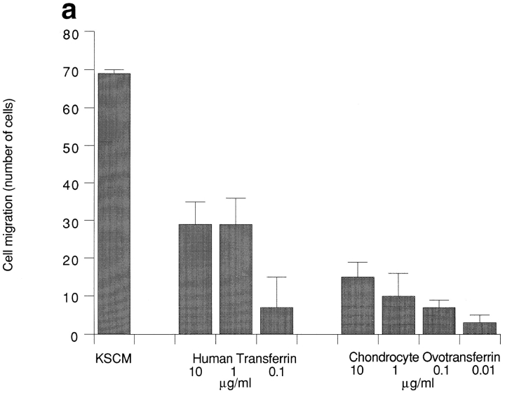
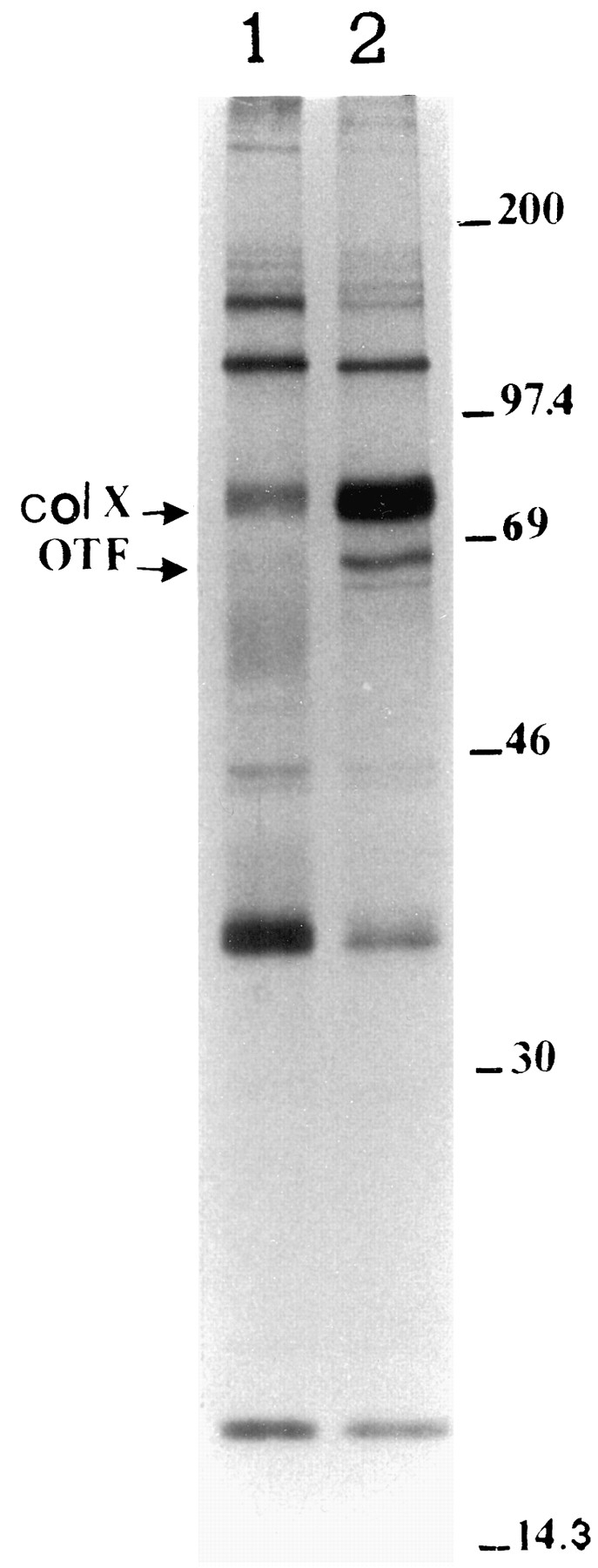
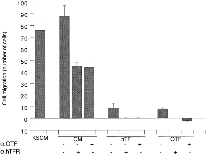
During endochondral bone formation, avascular cartilage differentiates to hypertrophic cartilage that then undergoes erosion and vascularization leading to bone deposition. Resting cartilage produces inhibitors of angiogenesis, shifting to production of angiogenic stimulators in hypertrophic cartilage. A major protein synthesized by hypertrophic cartilage both in vivo and in vitro is transferrin. Here we show that transferrin is a major angiogenic molecule released by hypertrophic cartilage. Endothelial cell migration and invasion is stimulated by transferrins from a number of different sources, including hypertrophic cartilage. Checkerboard analysis demonstrates that transferrin is a chemotactic and chemokinetic molecule. Chondrocyte-conditioned media show similar properties. Polyclonal anti-transferrin antibodies completely block endothelial cell migration and invasion induced by purified transferrin and inhibit the activity produced by hypertrophic chondrocytes by 50-70% as compared with controls. Function-blocking mAbs directed against the transferrin receptor similarly reduce the endothelial migratory response. Chondrocytes differentiating in the presence of serum produce transferrin, whereas those that differentiate in the absence of serum do not. Conditioned media from differentiated chondrocytes not producing transferrin have only 30% of the endothelial cell migratory activity of parallel cultures that synthesize transferrin. The angiogenic activity of transferrins was confirmed by in vivo assays on chicken egg chorioallantoic membrane, showing promotion of neovascularization by transferrins purified from different sources including conditioned culture medium. Based on the above results, we suggest that transferrin is a major angiogenic molecule produced by hypertrophic chondrocytes during endochondral bone formation.


Figures

Figure 1
Figure 1
Chemotactic (a) and chemoinvasive (b) responses of vascular EAhy 926 cells to human transferrin and chicken ovotransferrin purified from chondrocyte-conditioned medium. The factors were dissolved in F12 medium. F12 medium alone was used as control for background random migration that has been subtracted. Assays were performed in triplicate and repeated at least twice. Five fields were counted on each triplicate filter. Bars indicate SD. A typical experiment is shown. KSCM, control conditioned medium from Kaposi sarcoma cells.
Figure 1
Figure 1
Chemotactic (a) and chemoinvasive (b) responses of vascular EAhy 926 cells to human transferrin and chicken ovotransferrin purified from chondrocyte-conditioned medium. The factors were dissolved in F12 medium. F12 medium alone was used as control for background random migration that has been subtracted. Assays were performed in triplicate and repeated at least twice. Five fields were counted on each triplicate filter. Bars indicate SD. A typical experiment is shown. KSCM, control conditioned medium from Kaposi sarcoma cells.
Figure 2
Figure 2
Comparison of chemotactic responses of vascular cells to hypertrophic chondrocyte-conditioned culture medium, to human transferrin (hTF), to chicken egg white ovotransferrin (OTF), to ovotransferrin, purified from chondrocyte-conditioned medium (ChOTF), and to human bFGF. Conditioned medium (CM) was obtained from hypertrophic chondrocytes grown in suspension for ∼ 3 wk in the presence of ascorbic acid. F12 medium with 0.1% BSA was analyzed as control to verify background random migration. Assays were performed and analyzed as in Fig. 1 and repeated at least twice. Bars indicate SD. A typical experiment is shown.
Figure 3
Figure 3
Chemotactic (a) and chemoinvasive (b) responses of vascular cells to hypertrophic chondrocyte-conditioned culture medium and to ovotransferrin, purified from chondrocyte-conditioned medium, and their inhibition by affinity-purified antibodies against ovotransferrin. Conditioned medium (CM) was obtained from hypertrophic chondrocytes grown in suspension for ∼ 3 wk in the presence of ascorbic acid; ovotransferrin (OTF) was used at a concentration of 10 μg/ml. Analogous effects were obtained with a lower antibody concentration (10 μg/ml). Cell migration in the presence of antibodies alone in F12 was not significantly different from background random migration. Assays were performed and analyzed as in Fig. 1 and repeated at least twice on different culture media. Bars indicate SD. A typical experiment is shown. αOTF, experiments with antibodies against ovotransferrin at 50 μg/ml.
Figure 3
Figure 3
Chemotactic (a) and chemoinvasive (b) responses of vascular cells to hypertrophic chondrocyte-conditioned culture medium and to ovotransferrin, purified from chondrocyte-conditioned medium, and their inhibition by affinity-purified antibodies against ovotransferrin. Conditioned medium (CM) was obtained from hypertrophic chondrocytes grown in suspension for ∼ 3 wk in the presence of ascorbic acid; ovotransferrin (OTF) was used at a concentration of 10 μg/ml. Analogous effects were obtained with a lower antibody concentration (10 μg/ml). Cell migration in the presence of antibodies alone in F12 was not significantly different from background random migration. Assays were performed and analyzed as in Fig. 1 and repeated at least twice on different culture media. Bars indicate SD. A typical experiment is shown. αOTF, experiments with antibodies against ovotransferrin at 50 μg/ml.
Figure 4
Figure 4
Nonreducing 15% SDS-PAGE of metabolically labeled proteins from conditioned medium of hypertrophic chondrocytes, after 8 d of culture in suspension with ascorbic acid. (Lane 1) Medium not supplemented with FCS. (Lane 2) Medium supplemented with FCS. (Arrows) Ovotransferrin (OTF) and collagen type X. OTF was identified by immunoprecipitation (not shown).
Figure 5
Figure 5
Chemotactic migration induced in vascular cells by hypertrophic chondrocyte-conditioned media, after 9 d in suspension with ascorbic acid, with or without FCS, and inhibition by affinity-purified antibodies against ovotransferrin. A population of hypertrophic chondrocytes grown in suspension for 3 wk was split, and the suspension culture continued in the presence of ascorbic acid, either in the presence or absence of FCS. Conditioned media were collected after 9 d of suspension; for the assays, CM were diluted proportionally to the DNA content of the cells. Antibody concentration was 10 μg/ml. Cell migration in the presence of antibodies alone in F12 medium was not significantly different from background random migration. Assays were performed and analyzed as in Fig. 1 and repeated independently twice, on different culture media, with similar results. A typical experiment is shown. Bars indicate SD. FCS CM, conditioned medium from culture supplemented with FCS; SF CM, conditioned medium from serum-free cultures. αOTF, antibodies against ovotransferrin.
Figure 6
Figure 6
Western blot analysis for human transferrin receptor on EAhy 926 cells. Cells were scraped and lysed in 0.01% SDS in PBS. Aliquots of cell extracts were run on 15% SDS-PAGE in reducing conditions and blotted. The presence of the human transferrin receptor was detected with goat anti–human transferrin receptor antibodies. (Lane 1) negative control; (lane 2) anti–human transferrin receptor antibodies. (Arrow) Band with the expected 92-kD molecular mass of the transferrin receptor. (Left) Molecular mass standard migrations.
Figure 7
Figure 7
Chemotactic response by vascular cells and inhibition by the blocking mAb 42/6 against human transferrin receptor (αhTFR) and by polyclonal antibodies against ovotransferrin (αOTF). Chemoattractants were hypertrophic chondrocyte-conditioned culture medium, human transferrin, and ovotransferrin purified from chondrocyte-conditioned medium, as indicated. Antibody concentrations were 2.5 μg/ml (αhTFR) and 10 μg/ml (αOTF). Cell migration with antibodies alone in F12 was not significantly different from background random migration. Ovotransferrin (OTF) and human transferrin (hTF) were used at 10 μg/ml. Assays were performed and analyzed as in Fig. 1 and repeated at least twice on different culture media. A typical experiment is shown. Bars indicate SD. CM, conditioned medium from hypertrophic chondrocytes.
Figure 8
Figure 8
(A) CAM of 12-d-old chick embryo incubated for 4 d with a sponge adsorbed with 1 μg of ovotransferrin purified from chicken hypertrophic chondrocyte-conditioned medium. Note the presence of an increased number of blood vessels with a radially arranged “spoked wheel” pattern around the implant. (C) Vascular reaction is more detectable after India ink injection. (B and D) CAM of 12-d-old chick embryo incubated for 4 d with a sponge adsorbed with vehicle alone (PBS), used as negative control. No vascular response is detectable around the sponge in vivo (B) and after India ink injection (D). Bar, 330 μm.
Figure 9
Figure 9
(a) Histological section of a sponge treated with purified ovotransferrin. Note, among the sponge trabeculae, a collagenous matrix containing numerous capillaries (arrows) and a cellular infiltrate prevalently constituted by fibroblasts. (b) Histological section of a sponge treated with PBS. No collagenous matrix, blood vessels, and fibroblasts are detectable among the sponge trabeculae. Bar, 50 μm.

References

    1. Aizenman Y, Wechsel ME, de Vellis J. Changes in insulin and transferrin requirements of pure brain neuronal cultures during embryonic development. Proc Natl Acad Sci USA. 1986;83:2263–2266. - PMC - PubMed
    1. Albini A, Iwamoto Y, Kleinman HK, Martin GR, Aaronson SA, Kozlowski JM, McEwan RN. A rapid in vitro assay for quantitating the invasive potential of tumor cells. Cancer Res. 1987;4:3239–3245. - PubMed
    1. Albini A, Repetto L, Carlone S, Benelli R, Gendelman R, Filippi PD, Bussolino F, Monaco L, Soria M, Parravicini C. Characterization of Kaposi's sarcome-derived cell cultures from an epidemic and a classic case. Int J Oncol. 1992;1:723–730. - PubMed
    1. Albini A, Fontanini G, Masiello L, Tacchetti C, Bigini D, Luzzi P, Noonan DM, Stetler-Stevenson G. Angiogenic potential in vivo by Kaposi's sarcoma cell-free supernatants and HIV-1 tat product: inhibition of KS-like lesions by tissue inhibitor of metalloproteinase-2. AIDS (Phila) 1994;8:1237–1244. - PubMed
    1. Albini A, Benelli R, Presta M, Rusnati M, Ziche M, Rubartelli A, Paglialunga G, Bussolino F, Noonan D. HIV-tat protein is a heparinbinding angiogenic growth factor. Oncogene. 1996;12:289–297. - PubMed

Publication types

MeSH terms

Grants and funding