. 1997 Mar 17;185(6):1113-22.
doi: 10.1084/jem.185.6.1113.

An HLA-DR1 transgene confers susceptibility to collagen-induced arthritis elicited with human type II collagen

Affiliations
Free PMC article

An HLA-DR1 transgene confers susceptibility to collagen-induced arthritis elicited with human type II collagen

E F Rosloniec et al. J Exp Med. .
Free PMC article

Abstract

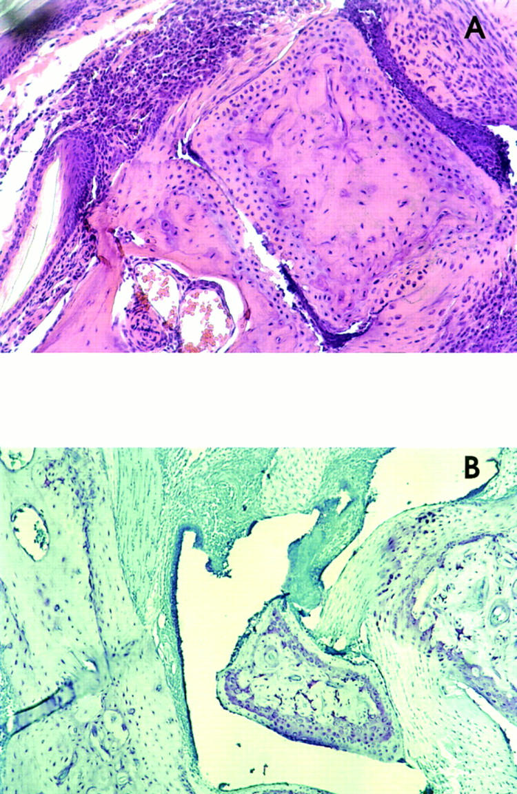
Rheumatoid arthritis (RA) is an autoimmune disease that is strongly associated with the expression of several HLA-DR haplotypes, including DR1 (DRB1*0101). Although the antigen that initiates RA remains elusive, it has been shown that many patients have autoimmunity directed to type II collagen (CII). To test the hypothesis that HLA-DR1 is capable of mediating an immune response to CII, we have generated transgenic mice expressing chimeric (human/mouse) HLA-DR1. When the DR1 transgenic mice were immunized with human CII (hCII), they developed a severe autoimmune arthritis, evidenced by severe swelling and erythema of the limbs and marked inflammation and erosion of articular joints. The development of the autoimmune arthritis was accompanied by strong DR1-restricted T and B cell responses to hCII. The T cell response was focused on a dominant determinant contained within CII(259-273) from which an eight amino acid core was defined. The B cell response was characterized by high titers of antibody specific for hCII, and a high degree of cross-reactivity with murine type II collagen. These data demonstrate that HLA-DR1 is capable of presenting peptides derived from hCII, and suggest that this DR1 transgenic model will be useful in the development of DR1-specific therapies for RA.


Figures

Figure 1
Figure 1
Expression of chimeric HLA-DR1 by lymphocytes from Tg mice. Two-color flow cytometry was performed to analyze the expression of DR1 and I-Af using mAbs L243 (anti-DR, PE labeled) and 103.6 (anti–I-Af, FITC labeled). In the DR1 Tg mice, virtually all of the cells that express I-Af, also express DR1. (A) Lymphocytes from Tg DR1 mice express both I-Af and DR1. (B) Lymphocytes from non-Tg B10.M mice expressing only I-Af. Contour plots are based on 5,000 cells analyzed.
Figure 2
Figure 2
HLA-DR1 expression confers susceptibility to CIA in DR1 Tg mice. In two separate experiments, mice were immunized subcutaneously at the base of the tail with 100 μg of human CII in CFA. (A) Mice (n = 7 per group) were boosted with 100 μg of human CII in incomplete Freund's adjuvant at 21 d. (B) Mice (n = 9 per group) were not boosted. Beginning at 3 wk after immunization, mice were monitored for inflammation of fore and hind paws. ▪, DR1 Tg mice; •, non-Tg mice.
Figure 3
Figure 3
Development of arthritis in DR1 Tg mice immunized with hCII. (A) Marked swelling of the tarsal and metatarsal joints, extending from the ankles through the digits of a DR1 Tg mouse immunized with hCII. (B) Normal hind paw of immunized non-Tg mouse. (C) Arthritic fore paw from DR1 Tg mouse. (D) Normal fore paw from an immunized non-Tg mouse.
Figure 4
Figure 4
Histopathology of arthritic joints from DR1 Tg mice immunized with hCII. (A) Hematoxylin and eosin stain of sections from an arthritic hind limb joint. Massive infiltration of inflammatory cells (upper left) and erosion of articular surfaces (lower articular joint) are clearly evident. (B) Toluidine blue stain of sections from an arthritic hind limb joint. Staining reveals proliferation of the synovial lining on the left side of the joint space as well as necrosis of the synovial lining (upper right portion of the joint). Samples were obtained 6 wk after hCII immunization.
Figure 5
Figure 5
Detection of antibody specific for hCII and mCII after immunization with hCII. Serum antibody specific for hCII and mCII was measured using solid phase ELISA as described in Materials and Methods. DR1 Tg mice (solid bars) produced high levels of antibody specific for both hCII and mCII in comparison to non-Tg control mice (open bars). A and B represent independent experiments and correspond respectively to A and B in Fig. 2. Sera were collected at 6 wk after immunization, and quantity of antibody was determined for individual mice. Data are expressed as mean relative units based on a standard antihCII sera. Error bars indicate the standard error of the mean.
Figure 6
Figure 6
Identification of the hCII immunodominant T cell determinants recognized by T cells from DR1 Tg and non-Tg mice. Mice were immunized with hCII and tested for their ability to respond to a panel of Mimotope peptides, 15 mer overlapping by 12 amino acids, spanning the entire length of the human α1(II) chain. The abscissa indicates the NH2-terminal residue number of the synthetic peptide. 10 μl of synthetic peptide (10–40 μg) was used in each proliferation assay. The data are expressed as DPM; (A) DR1 Tg mice, (B) nonTg mice. Mean [3H]thymidine incorporation in the absence of antigen was 3,696 DPM in A and 2,095 DPM in B. Data are representative of three independent experiments.
Figure 7
Figure 7
Identification of the core of the immunodominant determinant. hCII-primed T cells from DR1 Tg mice were tested for their ability to recognize a panel of Mimotope peptides, 15 mer, overlapping by 14 amino acids. Underlined residues indicate the deduced core of the T cell determinant. Data are expressed as DPM. T cell proliferation in the absence of antigen was 1,247 DPM.

References

    1. Stastny P, Ball EJ, Dry PJ, Nunez G. The human immune response region (HLA-D) and disease susceptibility. Immunol Rev. 1983;70:113–154. - PubMed
    1. Stastny P, Ball E, Kahn M, Olsen N, Pincus T, Gao X. HLA-DR4 and other genetic markers in rheumatoid arthritis. Br J Rheumatol. 1988;27:132–138. - PubMed
    1. Nepom GT, Byers P, Seyfried C, Healey LA, Wilske KR, Stage D, Nepom BS. HLA genes associated with rheumatoid arthritis. Identification of susceptibility alleles using specific oligonucleotide probes. Arthritis Rheum. 1989;32:15–21. - PubMed
    1. Wordsworth BP, Lanchbury JS, Sakkas LI, Welsh KI, Panayi GS, Bell JI. HLA-DR4 subtype frequencies in rheumatoid arthritis indicate that DRB1 is the major susceptibility locus within the HLA class II region. Proc Natl Acad Sci USA. 1989;86:10049–10053. - PMC - PubMed
    1. Gregersen P, Silver J, Winchester R. The shared epitope hypothesis: an approach to understanding the molecular genetics of susceptibility to rheumatoid arthritis. Arthritis Rheum. 1987;30:1205–1213. - PubMed

Publication types

MeSH terms