. 1997 Apr 15;17(8):2683-90.
doi: 10.1523/JNEUROSCI.17-08-02683.1997.

Signal transduction mediated by the truncated trkB receptor isoforms, trkB.T1 and trkB.T2

Affiliations

Signal transduction mediated by the truncated trkB receptor isoforms, trkB.T1 and trkB.T2

G T Baxter et al. J Neurosci. .

Abstract

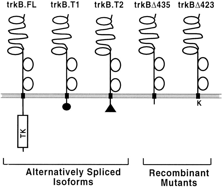
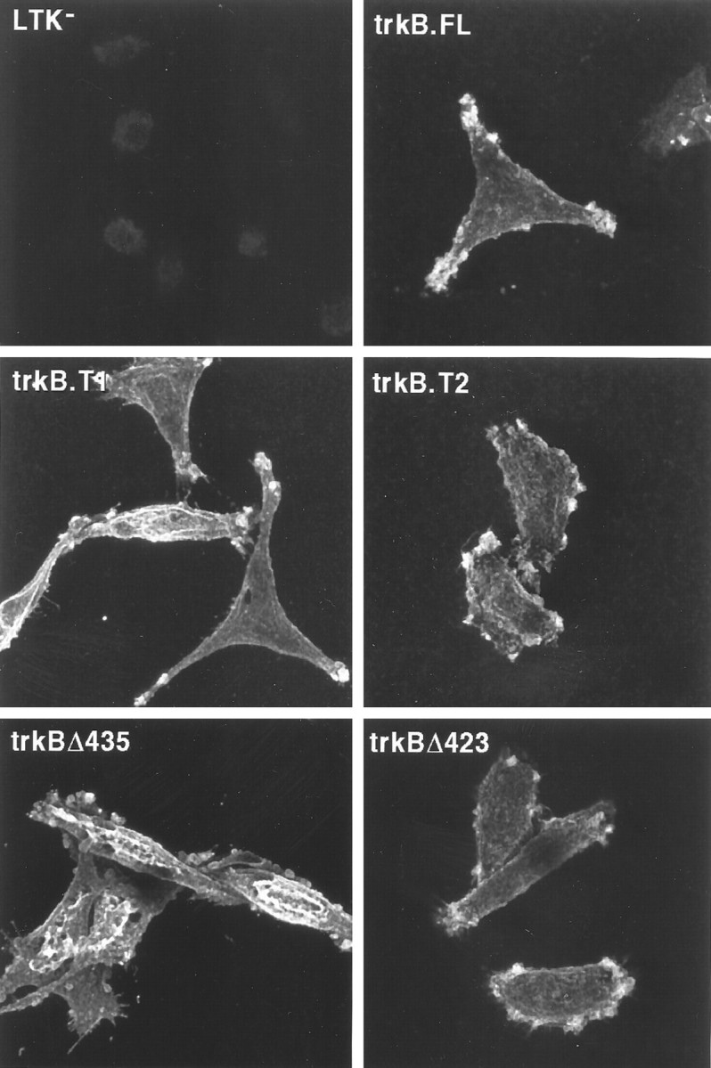
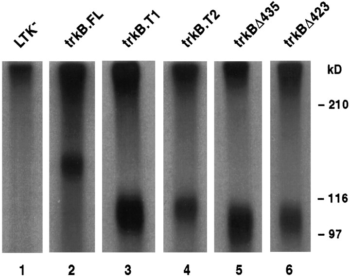
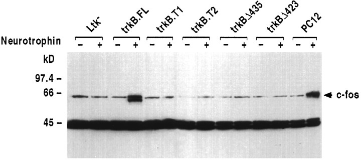
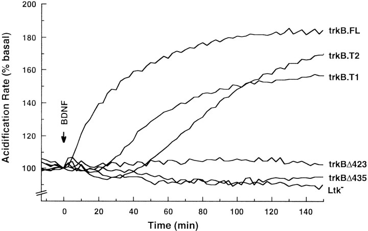
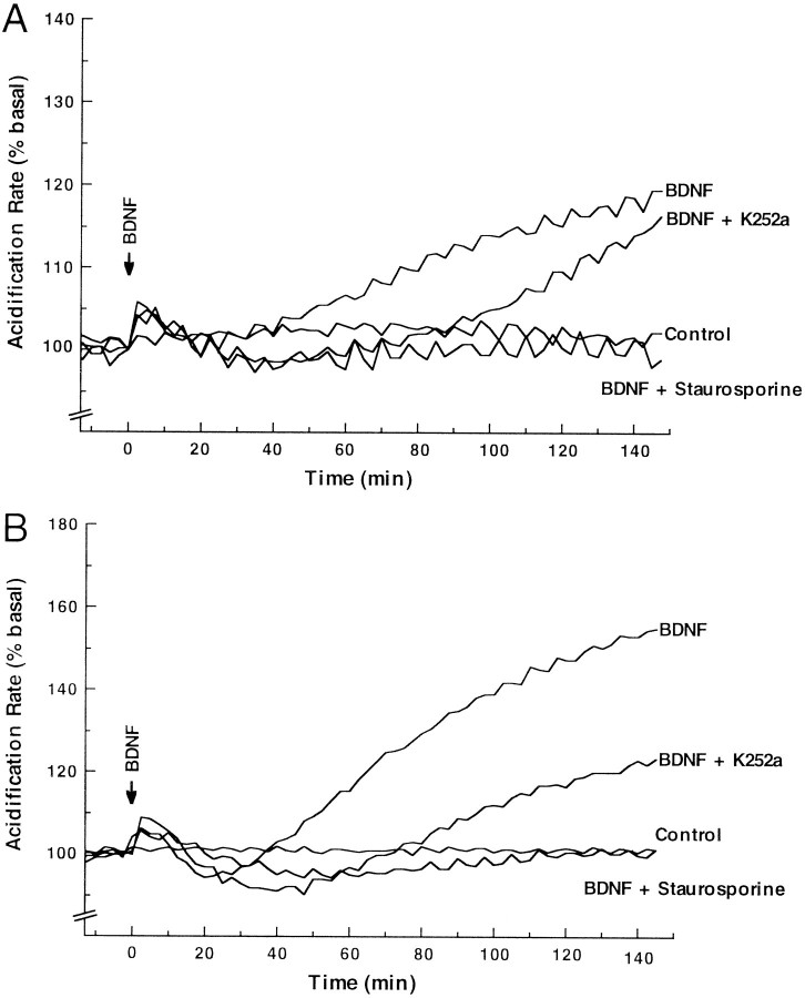
The trkB family of transmembrane proteins serves as receptors for BDNF and NT-4/5. The family is composed of a tyrosine kinase-containing isoform as well as several alternatively spliced "truncated receptors" with identical extracellular ligand-binding domains but very small intracellular domains. The two best-characterized truncated trkB receptors, designated as trkB.T1 and trkB.T2, contain intracellular domains of only 23 and 21 amino acids, respectively. Although it is known that the tyrosine kinase isoform (trkB.FL) is capable of initiating BDNF and NT-4/5-induced signal transduction, the functional role or roles of the truncated receptors remain enigmatic. At the same time, the potential importance of the truncated receptors in the development, maintenance, and regeneration of the nervous system has been highlighted by recent developmental and injury paradigm investigations. Here we have used trkB cDNA transfected cell lines to demonstrate that both trkB.T1 and trkB.T2 are capable of mediating BDNF-induced signal transduction. More specifically, BDNF activation of either trkB.T1 or trkB.T2 increases the rate of acidic metabolite release from the cell, a common physiological consequence of many signaling pathways. Further, these trkB.T1- and trkB. T2-mediated changes occur with kinetics distinct from changes mediated by trkB.FL, suggesting the participation of at least some unique rate-limiting component or components. Mutational analysis demonstrates that the isoform-specific sequences within the intracellular domains of each receptor are essential for signaling capability. Finally, inhibitor studies suggest that kinases are likely to be involved in the trkB.T1 and trkB.T2 signaling pathways.


Figures

Fig. 1.
Fig. 1.
Schematic diagram of wild-type and carboxy end-truncated trkB receptors used in this study.
Fig. 2.
Fig. 2.
Distribution of BDNF binding on the plasma membranes of wild-type (trkB.FL, trkB.T1, trkB.T2) and mutant (trkBΔ435 and trkBΔ423) trkB receptors in clonal, stably transfected L-cells: confocal microscopy. Transfected and nontransfected control cells were treated on coverslips with biotinylated BDNF, fixed, treated with streptavidin–Cy3, and viewed by laser scanning confocal microscopy. A Z-series of 0.5 μm optical sections encompassing the entire thickness of the cells was acquired; images presented here are a projection of all of the optical sections. Laser strength was 3% for trkB.T1, trkBΔ435, and trkBΔ423 transfected cells, and 10% for trkB.T2 and trkB.FLtransfected cells and for nontransfected control cells.
Fig. 3.
Fig. 3.
Biochemical characterization of cell-surface BDNF receptors on cells expressing wild-type (trkB.FL, trkB.T1, trkB.T2) and mutant (trkBΔ435 andtrkBΔ423) trkB receptors in clonal, stably transfected L-cells: 125I-BDNF cross-linking. Transfected and nontransfected control cells were treated with 125I-BDNF, cross-linked with EDAC, and lysed with Triton X-100 containing buffer. Then the lysate was enriched for trkB receptors by wheat germ agglutinin chromatography, fractionated by SDS-PAGE, and visualized by fluorography. Exposure times were 8 hr for trkB.T1 andtrkBΔ435 transfected cells, 3 d fortrkBΔ423 transfected cells, and 2 weeks fortrkB.T2 and trkB.FL transfected cells and for nontransfected control cells. (The x-ray film was not preflashed, which exaggerates the differences between stronger and weaker signals.)
Fig. 4.
Fig. 4.
trkB.FL transfectants, but nottrkB.T1, trkB.T2, trkBΔ435, ortrkBΔ423 transfectants, induce c-fos expression in response to BDNF administration. Cells were treated with 50 ng/ml recombinant BDNF for the indicated amounts of time; nuclei were isolated and immunoblotted with an anti-c-fos probe (Santa Cruz Biotechnology SC-52). The arrow marks the migration of the 62 kDa c-fos protein. PC12 cells were treated with 50 ng/ml NGF instead of BDNF, because they express trkA, but not trkB, receptors.
Fig. 5.
Fig. 5.
trkB.T1 and trkB.T2are both capable of mediating BDNF-dependent signal transduction requiring their respective intracellular domains. Transfected cell lines were treated with 50 ng/ml BDNF at time t = 0, and the rate of extracellular media acidification was monitored with a Cytosensor microphysiometer. Data are presented as the percentage of increase in acidification rate over initial basal activity before treatment with BDNF.
Fig. 6.
Fig. 6.
Effects of K252a and staurosporine on trkB.T1 and trkB.T2 signaling capability. A, TrkB.T1 transfected cell lines were pretreated for 20 min with either no inhibitor, 100 nm K252a, or 200 nm staurosporine and then treated simultaneously with the same inhibitor plus 50 ng/ml BDNF at time t = 0 and analyzed as in Figure 5.B, TrkB.T2 transfected cell lines were pretreated for 20 min with either no inhibitor, 100 nm K252a, or 200 nm staurosporine and then treated simultaneously with the same inhibitor plus 50 ng/ml BDNF at time t = 0 and analyzed as in Figure 5. All exposures to BDNF or BDNF plus inhibitor were for the duration of the experiment. For both the staurosporine and K252a experiments, these are relatively high concentrations that likely inhibit multiple kinases.

References

    1. Allendoerfer KL, Cabelli RJ, Escandón E, Kaplan DR, Nikolics K, Shatz CJ. Regulation of neurotrophin receptors during the maturation of the mammalian visual system. J Neurosci. 1994;14:1795–1811. - PMC - PubMed
    1. Armanini MP, McMahon SB, Sutherland J, Shelton DL, Philips HS. Truncated and catalytic isoforms of trkB are co-expressed in neurons of rat and mouse CNS. Eur J Neurosci. 1995;7:1403–1409. - PubMed
    1. Baxter GT, Miller DL, Kuo RC, Wada HG, Owicki JC. PKC epsilon is involved in granulocyte-macrophage colony-stimulating factor signal transduction: evidence from microphysiometry and antisense oligonucleotide experiments. Biochemistry. 1992;31:10950–10954. - PubMed
    1. Baxter GT, Young ML, Miller DL, Owicki JC. Using microphysiometry to study the pharmacology of exogenously expressed m1 and m3 muscarinic receptors. Life Sci. 1994;55:573–583. - PubMed
    1. Beck KD, Lamballe F, Klein R, Barbacid M, Schauwecker PE, McNeill TH, Finch CE, Hefti F, Day JR. Induction of noncatalytic TrkB neurotrophin receptors during axonal sprouting in the adult hippocampus. J Neurosci. 1993;13:4001–4014. - PMC - PubMed

Publication types

MeSH terms