

Transcranial magnetic stimulation during positron emission tomography: a new method for studying connectivity of the human cerebral cortex
- PMID: 9096152
- PMCID: PMC6573635
- DOI: 10.1523/JNEUROSCI.17-09-03178.1997
Transcranial magnetic stimulation during positron emission tomography: a new method for studying connectivity of the human cerebral cortex
Abstract
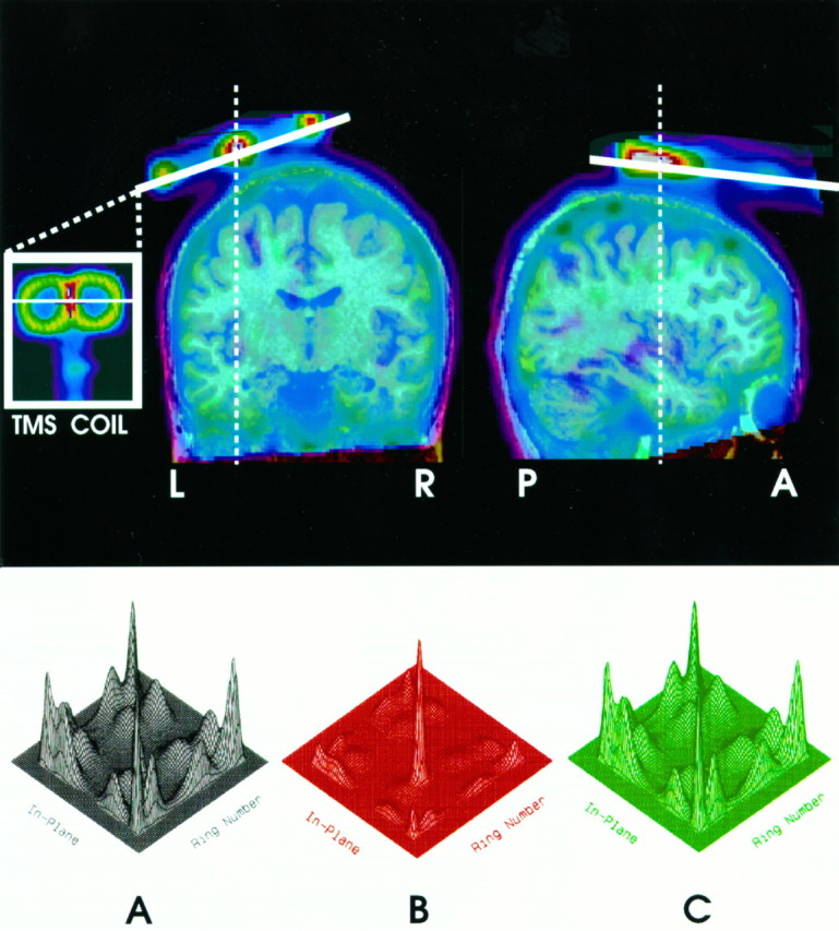
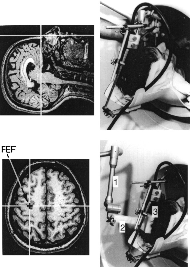
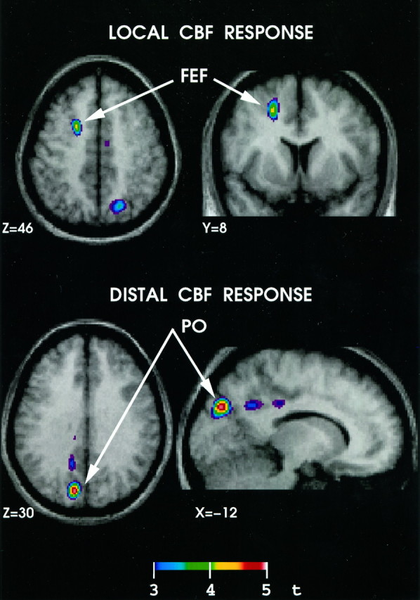
We describe a new technique permitting the mapping of neural connections in the living human brain. The method combines two well established tools of brain research: transcranial magnetic stimulation (TMS) and positron emission tomography (PET). We use TMS to stimulate directly a selected cortical area while simultaneously measuring changes in brain activity, indexed by cerebral blood flow (CBF), with PET. The exact location of the stimulation site is achieved by means of frameless stereotaxy. In the first study using this technique, we found significant positive correlations between CBF and the number of TMS pulse trains at the stimulation site, namely the left frontal eye field (FEF) and, most importantly, in the visual cortex of the superior parietal and medial parieto-occipital regions. The pattern of these distal effects was consistent with the known anatomic connectivity of the monkey FEF. We suggest that the combined TMS/PET technique offers an objective tool for assessing the state of functional connectivity without requiring the subject to engage in any specific behavior.
Figures
References
-
- Alexander GE, Moeler JR. Application of the scaled subprofile model to functional imaging in neuropsychiatric disorders: a principal component approach to modelling brain function in disease. Hum Brain Mapp. 1994;2:79–94.
-
- Amassian VE, Cracco RQ, Maccabee PJ, Cracco JB. Cerebello-frontal cortical projections in humans studied with the magnetic coil. Electroencephalogr Clin Neurophysiol. 1992;85:265–272. - PubMed
-
- Bailey P, von Bonin G, Garol HE, McCulloch WS. Long association fibers in cerebral hemispheres of monkey and chimpanzee. J Neurophysiol. 1943;6:129–134.
-
- Bailey P, von Bonin G, Davis EW, Garol HW, McCulloch WS. Further observations on associational pathways in the brain of Macaca mulatta. J Neuropathol Exp Neurol. 1944;3:413–415.
-
- Barker AT, Freeston IL, Jalinous R, Jarratt JA. A. Noninvasive stimulation of motor pathways within the brain using time-varying magnetic fields. Electroencephalogr Clin Neurophysiol. 1985;61:S245–S246.
Publication types
MeSH terms
LinkOut - more resources
Full Text Sources
Other Literature Sources