. 1997 Apr 1;94(7):2874-9.
doi: 10.1073/pnas.94.7.2874.

Crystal structure of varicella-zoster virus protease

Affiliations

Crystal structure of varicella-zoster virus protease

X Qiu et al. Proc Natl Acad Sci U S A. .

Abstract

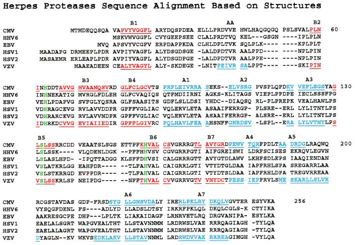
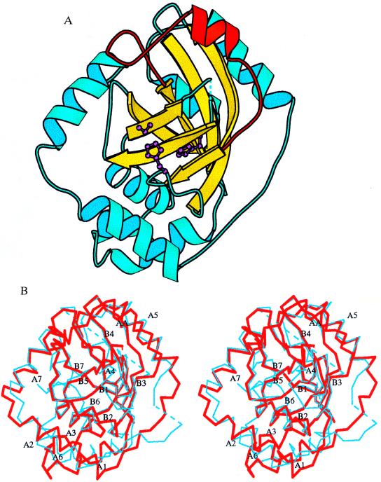
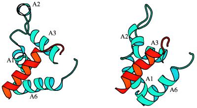
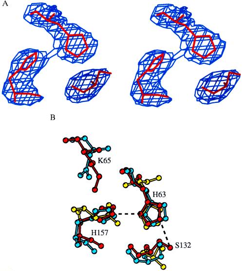
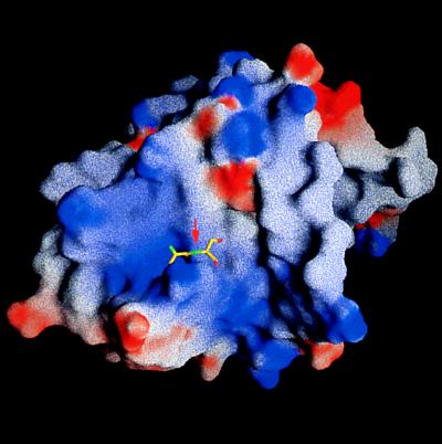
Varicella-zoster virus (VZV), an alpha-herpes virus, is the causative agent of chickenpox, shingles, and postherpetic neuralgia. The three-dimensional crystal structure of the serine protease from VZV has been determined at 3.0-A resolution. The VZV protease is essential for the life cycle of the virus and is a potential target for therapeutic intervention. The structure reveals an overall fold that is similar to that recently reported for the serine protease from cytomegalovirus (CMV), a herpes virus of the beta subfamily. The VZV protease structure provides further evidence to support the finding that herpes virus proteases have a fold and active site distinct from other serine proteases. The VZV protease catalytic triad consists of a serine and two histidines. The distal histidine is proposed to properly orient the proximal histidine. The identification of an alpha-helical segment in the VZV protease that was mostly disordered in the CMV protease provides a better definition of the postulated active site cavity and reveals an elastase-like S' region. Structural differences between the VZV and CMV proteases also suggest potential differences in their oligomerization states.


Figures

Figure 1
Figure 1
The structure-assisted alignment of human herpes virus proteases. The secondary structure elements of CMV and VZV protease are underlined and labeled. Helical (AA-A7) regions (blue), strands (B1-B7) (red), and the conserved catalytic triad (green). CMV numbering is used.
Figure 2
Figure 2
The monomeric structure of the VZV protease. (A) The core β-barrel (yellow), the catalytic triad (purple), and the AA loop (red). (B) Stereoview of the superposition between the VZV (thick red lines) and CMV (thin blue lines) protease structures. The secondary elements of the VZV protease are labeled. The diagram was drawn with the program molscript (19).
Figure 3
Figure 3
(Left) The dimer interface of VZV protease. (Right) CMV protease. Helix A6 of one of the monomers is shown in red; A1, A2, A3, and A6 of the other are shown in blue. The two A6 helices are not parallel to each other in VZV protease. The segments containing helices A2 assume quite different conformations in VZV and CMV proteases.
Figure 4
Figure 4
The catalytic residues (A) in the omit Fo − Fc map contoured at 3σ. (B) VZV protease (red) compared with CMV (blue) and trypsin (yellow). Dashed lines connects the catalytic triad of the VZV protease. Only CMV numbering is used.
Figure 5
Figure 5
Molecular surface of the VZV protease looking into the postulated substrate binding groove. The surface is color-coded by electrostatic potentials (blue for positive and red for negative) calculated with the program grasp (28). Modeled is the Ala-Ser cleavage site. The red arrow indicates the position of the scissile bond.

References

    1. Straus S E. Ann Neurol. 1994;35:S11–S12. - PubMed
    1. Gao M, Matusick-Kumar L, Hurlburt W, DiTusa S F, Newcomb W W, Brown J C, McCann P J, III, Deckman I, Colonno R J. J Virol. 1994;68:3702–3712. - PMC - PubMed
    1. Liu F, Roizman B. J Virol. 1991;65:5149–5156. - PMC - PubMed
    1. Liu F, Roizman B. J Virol. 1993;67:1300–1309. - PMC - PubMed
    1. Liu F, Roizman B. Proc Natl Acad Sci USA. 1992;89:2076–2080. - PMC - PubMed

Substances

Associated data

LinkOut - more resources