. 1997 Apr 1;94(7):2975-80.
doi: 10.1073/pnas.94.7.2975.

Crystal structure of desheptapeptide(B24-B30)insulin at 1.6 A resolution: implications for receptor binding

Affiliations

Crystal structure of desheptapeptide(B24-B30)insulin at 1.6 A resolution: implications for receptor binding

S J Bao et al. Proc Natl Acad Sci U S A. .

Abstract

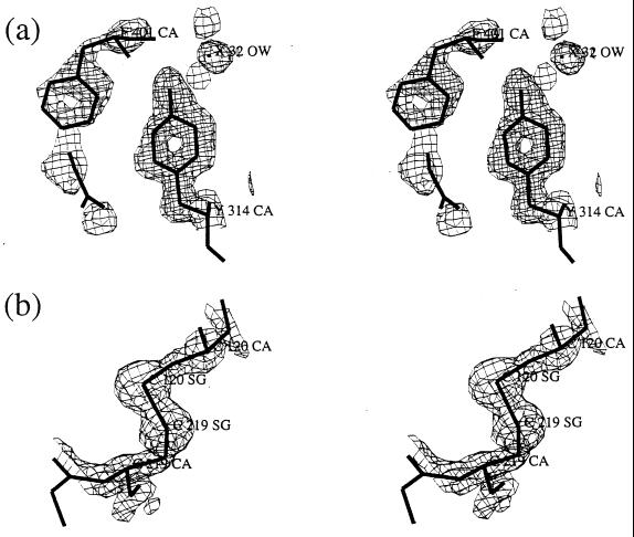
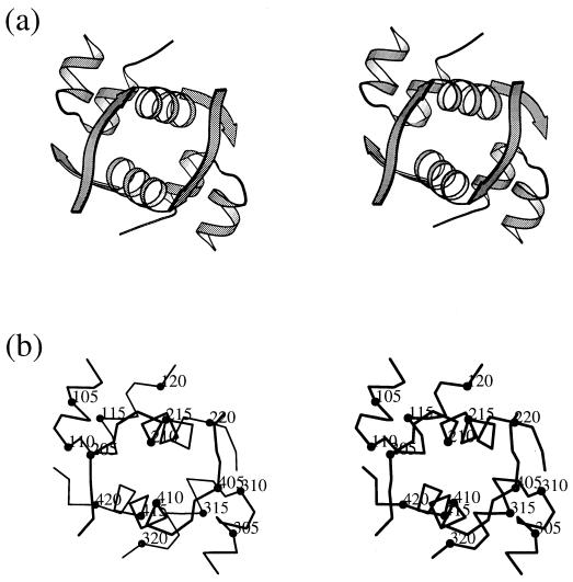
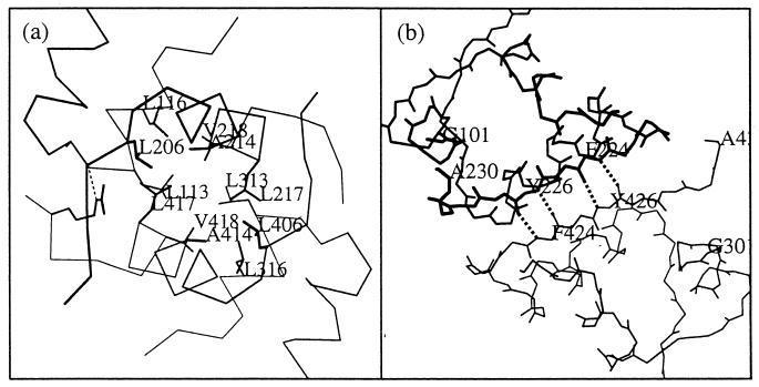
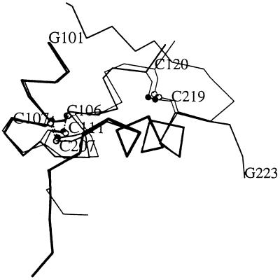
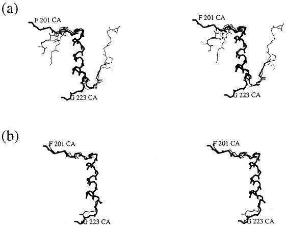
The crystal structure of desheptapeptide (B24-B30) insulin (DHPI), a virtually inactive analog of insulin, was determined at 1.6 A resolution. In the refined structure model, DHPI retains three alpha-helices (A1-A8, A12-A18, and B9-B19) as its structural framework, while great conformational changes occur in the N and C termini of B-chain. The beta-turn, which lies in B20-B30 in insulin and insulin analogs with high potency, no longer exists in DHPI. Relative motion is observed among the three alpha-helices, each as a rigid functional group. In contrast, a region covering B5-B6 and A6-A11 exhibits a relatively stable conformation. We interpret our results as identifying: (i) the importance of beta-turn in determining the receptor-binding potency of insulin and (ii) a leading role of PheB24 in maintaining the beta-turn structure.


Figures

Figure 1
Figure 1
Representative electron density. The stereoviews show: (a) the 2FoFc maps of PheB1 of molecule 2 and its nearby area, contoured at 1.0 σ, and (b) the S—S bond between A20 and B19 of molecule 1, contoured at 2.0 σ. The residues are labeled as follows: A-chain, molecule 1, 101–121; B-chain, molecule 1, 201–223; A-chain, molecule 2, 301–321; and B-chain, molecule 2, 401–423. [This figure was drawn with the program frodo (27).]
Figure 2
Figure 2
Ramachandran plot of DHPI structure. Glycine residues are shown as rectangles. [This figure was drawn with the program frodo (27).]
Figure 3
Figure 3
The overall structure of DHPI. (a) A stereo ribbon diagram of DHPI showing view along the local 2-fold axis near (0,0,Z). (b) Stereo Cα trace of two DHPI molecules, with every fifth residue labeled in each chain. Residues are labeled as in Fig. 1. [This figure was produced using molscript (28).]
Figure 4
Figure 4
Contacts of two monomers of DHPI or 2Zn insulin in an asymmetric unit. Molecule 1 of DHPI or 2Zn insulin is shown in boldface while molecule 2 is shown in regular type. (a) Contacts through hydrophobic interfaces in two DHPI monomers. The residues involved in the hydrophobic interaction are highlighted in boldface lines. A hydrogen bond linking Cys111-O and Arg422-NE is shown with a dotted line. (b) Contacts through β-strands of C-terminal B-chains in 2Zn insulin dimer. The hydrogen bonds are shown with dotted lines, with bonding residues labeled. Residues are labeled as in Fig. 1. [This figure was produced using molscript (28).]
Figure 5
Figure 5
Relative motion among the α-helices and S—S bonds. DHPI molecule 1 (boldface lines) and 2Zn insulin molecule 1 (regular lines) are superposed in main chains of B9–B19. The sulfur atoms are shown as circles while the S—S bonds are indicated with dotted lines. Residues are labeled as in Fig. 1. [This figure was produced using molscript (28).]
Figure 6
Figure 6
Structural comparison of DHPI with insulin and insulin analogs. (a) Main chain superposition of B-chains of DHPI molecule 1 (boldface lines) and 2Zn insulin, DPI, DHI, and des (B1–B2) despentapeptide (B26–B30) insulin (regular lines). Only B9–B19 were used to calculate the superposition matrices. (b) Similar comparison of DHPI molecule 1 (boldface lines) and molecule 2 (regular lines). Residues are labeled as in Fig. 1. [This figure was drawn with the program frodo (27).]

References

    1. Olefsky J, Saekow M, Tager H, Rubenstein A. J Biol Chem. 1980;255:6098–6105. - PubMed
    1. Tager H, Thomas N, Assoian R, Rubenstein A, Saekow M, Olefsky J, Kaiser E T. Proc Natl Acad Sci USA. 1980;77:3181–3185. - PMC - PubMed
    1. Shoelson S, Haneda M, Blix P, Nanjo A, Sanke T, Inouye K, Steiner D, Rubenstein A, Tager H. Nature (London) 1983;302:540–543. - PubMed
    1. Wang C C, Liang D C. Chin Biochem J. 1985;1:9–12.
    1. Liang D C, Chang W R, Zhang J P, Wan Z L. Sci China Ser B. 1992;35:547–557. - PubMed

Publication types

Associated data