

doi: 10.1073/pnas.94.7.2993.
Local DNA stretching mimics the distortion caused by the TATA box-binding protein
Affiliations
- PMID: 9096334
- PMCID: PMC20310
- DOI: 10.1073/pnas.94.7.2993
Item in Clipboard
Local DNA stretching mimics the distortion caused by the TATA box-binding protein
Proc Natl Acad Sci U S A.
.
Abstract
X-ray structures of the TATA box-binding protein complexed with its DNA target show that the nucleic acid is severely bent away from the protein and also strongly unwound. We have used molecular mechanics and energy mapping to understand how such an unusual conformation can be induced. The results show that simple deformation pathways involving local stretching or unwinding of DNA reproduce many features of the experimental structure. Notably, kinked junctions with the flanking B-DNA regions occur without the need for any specific local
Figures
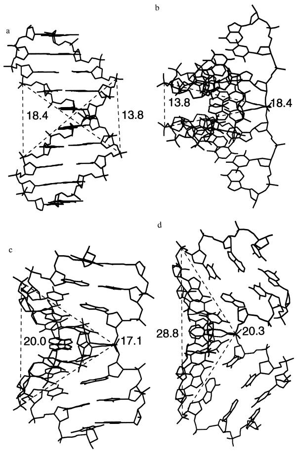
Interstrand distances between the
phosphorus atoms flanking a TATA sequence within DNA.
(a) B-DNA viewed perpendicularly to the helical axis.
(b) B-DNA viewed along the 5′–5′ vector.
(c) A-DNA viewed along the 5′–5′ vector.
(d) The complexed TATA box (1) viewed along the 5′–5′
vector.
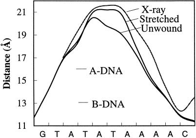
The 3′–3′ phosphorus distances (Å) for
successive dinucleotide base pairs along the TATA box sequence for the
x-ray structure of the TATA box (1) and the simulated stretched and
unwound conformations (the curve shown is a polynomial fit to the
successive P–P distances). The values for canonical A- and B-DNA are
shown for comparison.
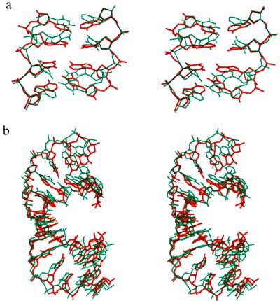
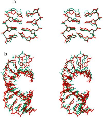
Superposition of the x-ray structure of the
complexed TATA box (red) with the stretched model conformation (blue)
of the (AT)n alternating oligomer for (a)
the central TATA sequence and (b) the 12 bp of the bound
duplex. The upper view is into the minor groove.
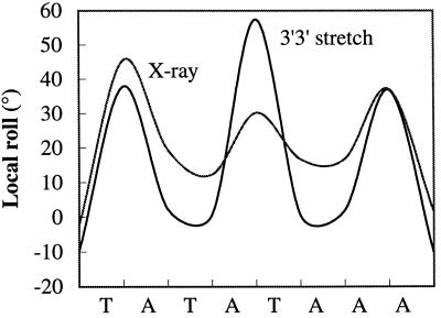
Comparison of the local roll angles along the
TATA box sequence for the x-ray conformation (1) and for the stretched
model (AT)n oligomer (the curve shown is a polynomial fit
to the successive roll angles).
Superposition of the x-ray structure of the
complexed TATA box (red) with the stretched model conformation of an
oligomer with an identical base sequence (blue) for (a)
the central TATA sequence and (b) the 12 bp of the bound
duplex. The upper view is into the minor groove.
References
-
- Kim Y, Gieger J H, Hahn S, Sigler P B. Nature (London) 1993;365:512–520. - PubMed
-
- Kim J L, Nikolov D B, Burley S K. Nature (London) 1993;365:520–527. - PubMed
-
- Kim J L, Burley S K. Nat Struct Biol. 1994;1:638–653. - PubMed
-
- Tan S, Hunziker Y, Sargent D F, Richmond T J. Nature (London) 1996;381:127–134. - PubMed
-
- Geiger J H, Hahn S, Lee S, Sigler P. Science. 1996;272:830–836. - PubMed
Publication types
MeSH terms
Substances
LinkOut - more resources
Full Text Sources
