. 1997 Apr 1;94(7):3016-21.
doi: 10.1073/pnas.94.7.3016.

Identification of a novel pathway important for proliferation and differentiation of primary erythroid progenitors

Affiliations

Identification of a novel pathway important for proliferation and differentiation of primary erythroid progenitors

U Klingmüller et al. Proc Natl Acad Sci U S A. .

Abstract

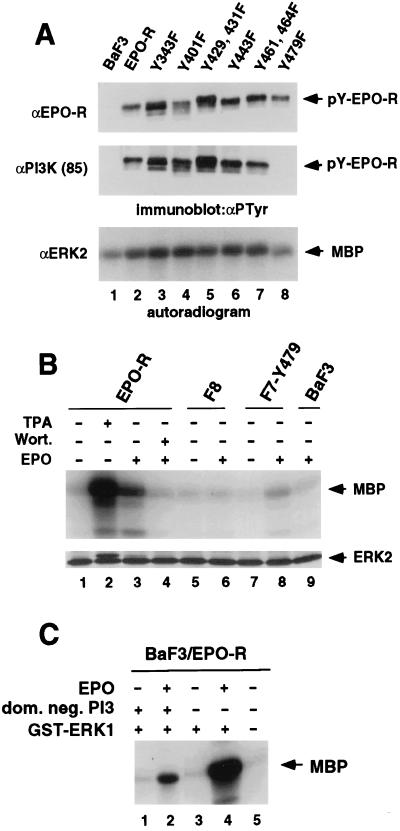
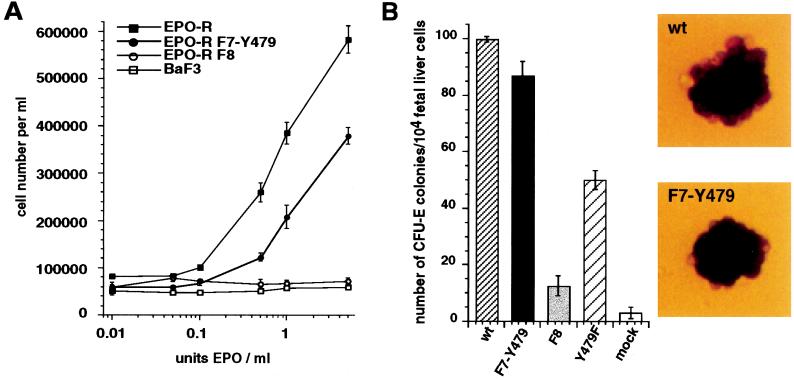
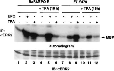
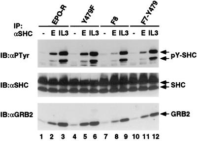
Homodimerization of the erythropoietin (EPO) receptor (EPO-R) in response to EPO binding transiently activates the receptor-associated protein tyrosine kinase JAK2. Tyrosine phosphorylation of the EPO-R creates "docking sites" for SH2 domain(s) in signaling molecules such as the protein tyrosine phosphatases SH-PTP1 and SH-PTP2, phosphoinositide 3-kinase (PI3 kinase), and STAT5. However, little is known about the specific intracellular signals essential for proliferation and differentiation of erythroid progenitors. Here we show that an EPO-R containing only one cytosolic (phospho)tyrosine residue, Y479, induces a signal transduction pathway sufficient for proliferation and differentiation of fetal liver progenitors of erythroid colony-forming units from EPO-R(-/-) mice as well as for proliferation of cultured hematopoietic cells. This cascade involves sequential EPO-induced recruitment of PI3 kinase to the EPO-R and activation of mitogen-activated protein kinase activity, independent of the Shc/Grb2-adapter pathway and of STAT5. Protein kinase C epsilon may be one of the mediators connecting PI3 kinase with the mitogen-activated protein kinase signaling cascade. Our results identify a signaling cascade important in vivo for erythroid cell proliferation and differentiation.


Figures

Figure 2
Figure 2
Tyr-479 is essential for association of the EPO-R with the p85 subunit of PI3 kinase and is important for MAPK activation. (A) Parental BaF3 cells and BaF3 cells expressing the wild-type EPO-R or the various mutant EPO-Rs were stimulated with EPO, lysed, and used for immunoprecipitation with antisera directed against the EPO-R, the regulatory subunit of PI3 kinase (p85), or ERK2 as indicated. The tyrosine phosphorylated form of the immunoprecipitated receptor (pY-EPO-R) was detected by immunoblotting with the monoclonal anti-PTyr antibody 4G10 (αPTyr) and is indicated by arrows (Top and Middle). MAPK immunoprecipitated was subjected to an in vitro kinase assay using [γ-32P]ATP. Phosphorylation of the exogenous substrate MBP was detected by autoradiography and is marked by an arrow (Bottom). (B) Tyr-479 is sufficient for activation of MAPK. Prior to the MAPK assays, parental BaF3 cells and BaF3 cells expressing the wild-type EPO-R or mutant EPO-Rs F8 or F7Y479 were starved as described in Materials and Methods and either left unstimulated (−) or treated for 5 min with 100 units EPO per ml (+) prior to lysis. Stimulation for 15 min with 200 nM TPA in the absence of EPO and pre treatment of cells with 100 nM wortmannin in the presence of EPO are indicated (+). MAPK was immunoprecipitated by an antiserum directed against ERK2 and subjected to an in vitro kinase assay using [γ-32P]ATP. Phosphorylation of the exogenous substrate MBP was detected by autoradiography and equal immunoprecipitation of the ERK2 MAPK was confirmed by immunoblotting with anti-ERK2. The electrophoretic mobility shift of ERK2 detected in lane 2 and faintly in lanes 3 and 8 indicates the activation of MAPK due to phosphorylation. Similar results (not shown) were obtained when an antibody directed against ERK1 was used for immunoprecipitation. (C) An expression vector encoding a GST-ERK1 fusion protein (+) was transiently cotransfected either with an expression vector encoding a dominant negative form of PI3 kinase (dom. neg. PI3) (+) or a control vector into BaF3 cells expressing the EPO-R (BaF3/EPO-R). Twenty-four hours after transfection cells were starved for 4 hr as described and either left unstimulated (−) or treated for 5 min with 100 units EPO per ml (+). The GST-ERK1 fusion protein was precipitated from the lysates by absorption to glutathione agarose beads (Sigma) and subjected to an in vitro MAPK assay as described in Materials and Methods. Each transfection was done in triplicate and the autoradiogram of one representative experiment is shown.
Figure 1
Figure 1
Tyr-479 in the EPO-R is sufficient to promote EPO-dependent proliferation and differentiation. (A) Proliferation of parental BaF3 cells and BaF3 cells expressing the EPO-R or the mutant EPO-Rs F8 or F7Y479 in different concentrations of EPO. The number of cells was determined using a Coulter counter. The average cell numbers per milliliter in four independent determinations (±SD) are plotted against the concentration of EPO (units/ml) added. (B) Tyr-479 in the EPO-R is sufficient to support EPO-dependent differentiation of erythroid progenitors. Fetal liver cells harvested from day 12.5 EPO-R−/− mice were infected with retroviruses designed to express either the wild-type EPO-R or the point mutant EPO-Rs F7Y479, F8, or Y479F. The number of benzidine-positive colonies (CFU-Es) formed in the presence of EPO and stem cell factor was counted after 2 days. The results plotted represent the mean (±SD) of three independent experiments. All values are expressed per 104 nucleated fetal liver cells. The morphology of CFU-E colonies formed in the presence of EPO and stem cell factor in cultures infected with retroviruses expressing the wild-type EPO-R (wt) or the mutant EPO-R F7Y479 (F7-Y479) is the same.
Figure 3
Figure 3
Stimulation of MAPK activity mediated by the EPO-R involves recruitment of PI3 kinase but does not require maximal activation of PKCα. BaF3 cells expressing the wild-type EPO-R or the mutant EPO-R F7Y479 were either left untreated or were pretreated with TPA as described in A, indicated by + TPA (18 h). Prior to lysis cells were left unstimulated (−) or were stimulated either for 5 min with 100 units EPO/ml (+) or for 10 min with 1 μg/ml TPA (+). MAPK activity was immunoprecipitated using an antiserum directed against ERK2 (IP: αERK2) and subjected to an in vitro kinase assay in the presence of MBP and [γ-32P]ATP as described. MBP-phosphorylation was detected by analysis on a 15% SDS/PAGE, transfer to a nitrocellulose membrane, and autoradiography (Upper). Immunoblotting with an antiserum against ERK2 (IB: αERK2) demonstrates equal loading in all lanes (Lower).
Figure 4
Figure 4
Stimulation of MAPK activity mediated by the EPO-R does not require maximal activation of the Shc/Grb2-Adapter pathway. After starvation, BaF3 cells expressing the wild-type EPO-R or the mutant EPO-Rs Y479F, F8, or F7Y479 were left unstimulated or stimulated with either 100 units EPO/ml (E) or 1 μg/ml interleukin 3. Lysates were subjected to immunoprecipitation with 5 μg polyclonal rabbit serum directed against Shc. Analysis of immunoprecipitated proteins was conducted by separation on a 15% low-bis SDS/PAGE and successive immunoblotting with anti-PTyr, anti-Shc, and anti-Grb2 antibodies. The positions of tyrosine-phosphorylated Shc (pY-Shc), Shc, and Grb2 are indicated by arrows.

References

    1. D’Andrea A D, Lodish H F, Wong G G. Cell. 1989;57:277–285. - PubMed
    1. Watowich S S, Yoshimura A, Longmore G D, Hilton D J, Yoshimura Y, Lodish H F. Proc Natl Acad Sci USA. 1992;89:2140–2144. - PMC - PubMed
    1. Wrightom N C, Farrell F X, Chang R, Kashyap A K, Barbone F P, Mulcahy L S, Johnson D L, Barrett R W, Jolliffe L K, Dower W J. Science. 1996;273:397–402. - PubMed
    1. Witthuhn B A, Quelle F W, Silvennoinen O, Yi T, Tang B, Miura O, Ihle J N. Cell. 1993;74:227–236. - PubMed
    1. Klingmüller U, Lorenz U, Cantley L C, Neel B G, Lodish H F. Cell. 1995;80:729–738. - PubMed

Publication types

MeSH terms