Case Reports
. 1997 Apr 1;94(7):3116-21.
doi: 10.1073/pnas.94.7.3116.

A common mutational pattern in Cockayne syndrome patients from xeroderma pigmentosum group G: implications for a second XPG function

Affiliations
Case Reports

A common mutational pattern in Cockayne syndrome patients from xeroderma pigmentosum group G: implications for a second XPG function

T Nouspikel et al. Proc Natl Acad Sci U S A. .

Retraction in

Abstract

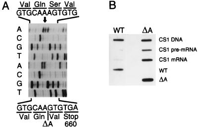
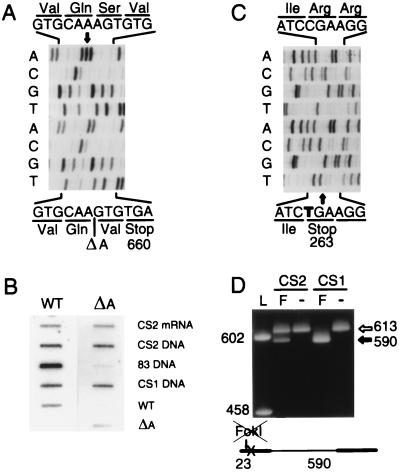
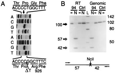
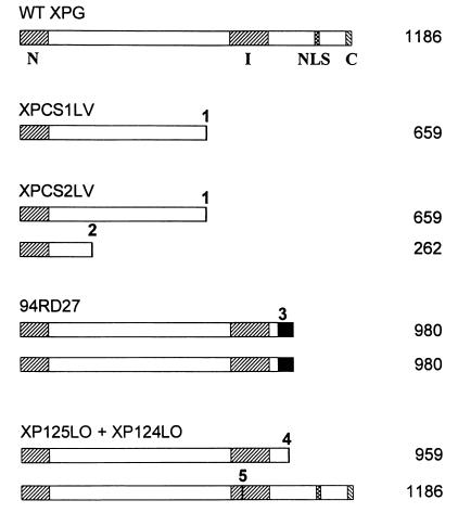
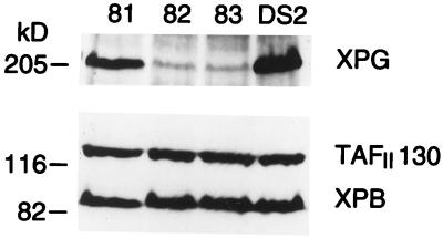
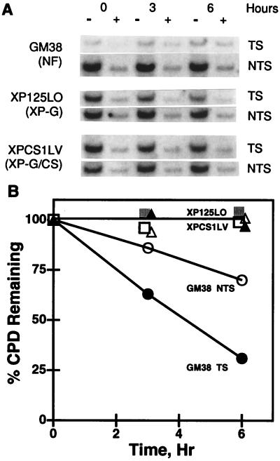
Xeroderma pigmentosum (XP) patients have defects in nucleotide excision repair (NER), the versatile repair pathway that removes UV-induced damage and other bulky DNA adducts. Patients with Cockayne syndrome (CS), another rare sun-sensitive disorder, are specifically defective in the preferential removal of damage from the transcribed strand of active genes, a process known as transcription-coupled repair. These two disorders are usually clinically and genetically distinct, but complementation analyses have assigned a few CS patients to the rare XP groups B, D, or G. The XPG gene encodes a structure-specific endonuclease that nicks damaged DNA 3' to the lesion during NER. Here we show that three XPG/CS patients had mutations that would produce severely truncated XPG proteins. In contrast, two sibling XPG patients without CS are able to make full-length XPG, but with a missense mutation that inactivates its function in NER. These results suggest that XPG/CS mutations abolish interactions required for a second important XPG function and that it is the loss of this second function that leads to the CS clinical phenotype.


Figures

Figure 1
Figure 1
Mutations in the XPG gene of patient XPCS1LV. (A) Sequence of wild-type (top 4 rows) and XPCS1LV (bottom 4 rows) RT-PCR products showing the single nucleotide deletion within the AAA triplet at nucleotides 2170–2172. (B) Slot-blot hybridization of PCR products with wild-type (WT) or mutation-specific (ΔA) oligonucleotide probes. The wild-type sequence is present in genomic DNA from XPCS1LV, but not in pre-mRNA or mRNA. cDNA clones WT and ΔA are included as controls.
Figure 2
Figure 2
Mutations in the XPG gene of patient XPCS2LV. (A and C) Sequence of wild-type (top) and XPCS2LV (bottom) RT-PCR products showing the same A deletion as in XPCS1LV in one allele (A) and the C → T transition at position 984 in the second allele (C). (B) Slot-blot hybridization of PCR products with wild-type (WT) or mutation-specific (ΔA) oligonucleotide probes. The wild-type and mutant sequences are present in both genomic DNA and mRNA from XPCS2LV. Genomic DNA from XPCS1LV and XP125LO (83 DNA) and cDNA clones WT and ΔA are included as controls. (D) FokI restriction analysis of genomic PCR products from XPCS2LV (CS2) and XPCS1LV (CS1). L, HaeIII digest of pBluescript II-SK+ DNA, with fragment lengths in bp; F, FokI digests; −, undigested products. The C → T transition destroys a FokI site (GGATG[N]9/and/[N]13CATCC) in one allele of XPCS2LV.
Figure 3
Figure 3
Mutations in the XPG gene of patient 94RD27. (A) Sequence of wild-type (top) and 94RD27 (bottom) RT-PCR products showing the T deletion at position 2972. (B) NciI restriction analysis of RT-PCR and genomic PCR products from RD9427 (lanes 94) and a wild-type control (Ctrl). −, Undigested products; N, NciI digests; L, HaeIII digest of pBluescript II-SK+ DNA, with fragment lengths in bp. The T deletion creates a new NciI site (CC/SGG) that is absent from control RNA and DNA. Note that the uncut 94RD27 genomic PCR product is slightly shorter than the control and that it is fully cut by NciI to yield 57-bp and 42-bp fragments.
Figure 4
Figure 4
Alignment of wild-type XPG with those predicted in the three XP-G/CS patients and the two sibling non-CS XP-G patients. The number of amino acids in each protein is listed on the right. Hatched areas indicate: N and I, conserved regions; NLS, putative bipartite nuclear localization signal; C, basic domain. Mutations: 1, Single A deletion within nucleotides 2170–2172; 2, nonsense C → T transition at nucleotide 984; 3, single T deletion at position 2972 (black area indicates the frameshift between amino acids 926 and 980); 4, nonsense G → T transversion at nucleotide 3075; and 5, missense C → T transition at nucleotide 2572.
Figure 5
Figure 5
Western blot analysis of lymphoblast extracts probed with antibodies to XPG, TAFII130, and XPB. The lymphoblast lines XPG82 (lane 82) and XPG83 (lane 83) are derived from patients XP124LO and XP125LO, respectively; XPG81 (lane 81) is from their obligate heterozygote mother. DS2 is a wild-type lymphoblast line (40). Note that the XPG protein, which has a predicted molecular mass of 133 kDa, is present in the two XP-G patients, but in reduced amounts, and that it migrates very close to the 205-kDa myosin marker.
Figure 6
Figure 6
Strand-selective repair of CPDs in the MTIA gene. (A) Human primary fibroblasts were exposed to 10 J/m2 UV and were allowed to repair for the indicated times. EcoRI-digested genomic DNA was treated (+) or not (−) with T4 endonuclease V, electrophoresed, blotted, and hybridized with RNA probes that distinguish the transcribed (TS) and nontranscribed (NTS) strands of the MTIA gene. (B) Repair values calculated from scanning densitometry measurements of the hybridization signals in the autoradiogram. The mean of all six 0 time values, 0.71 ± 0.03 (SD) CPD per 10 kb, was set equal to 100%. Filled symbols represent TS; open symbols represent NTS. Circles, normal fibroblasts, GM38; squares, XP-G fibroblasts XP125LO; and triangles, XP-G/CS fibroblasts, XPCS1LV.

Comment in

  • Findings of scientific misconduct.
    [No authors listed] [No authors listed] NIH Guide Grants Contracts (Bethesda). 2006 Jun 9:NOT-OD-06-075. NIH Guide Grants Contracts (Bethesda). 2006. PMID: 16764107 Free PMC article. No abstract available.
  • Findings of Scientific Misconduct.
    [No authors listed] [No authors listed] Fed Regist. 2006 Jun 8;71(110):33308-33309. Fed Regist. 2006. PMID: 27737147 Free PMC article. No abstract available.

References

    1. Friedberg E C, Walker G C, Siede W. DNA Repair and Mutagenesis. Washington, DC: Am. Soc. Microbiol.; 1995.
    1. Aboussekhra A, Biggerstaff M, Shivji M K K, Vilpo J A, Moncollin V, Podust V N, Protic M, Hübscher U, Egly J-M, Wood R D. Cell. 1995;80:859–868. - PubMed
    1. Mu D, Hsu D S, Sancar A. J Biol Chem. 1996;271:8285–8294. - PubMed
    1. Nance M A, Berry S A. Am J Med Genet. 1992;42:68–84. - PubMed
    1. Mellon I, Spivak G, Hanawalt P C. Cell. 1987;51:241–249. - PubMed

Associated data