. 1997 Apr 1;94(7):3352-6.
doi: 10.1073/pnas.94.7.3352.

HCAR and MCAR: the human and mouse cellular receptors for subgroup C adenoviruses and group B coxsackieviruses

Affiliations

HCAR and MCAR: the human and mouse cellular receptors for subgroup C adenoviruses and group B coxsackieviruses

R P Tomko et al. Proc Natl Acad Sci U S A. .

Abstract

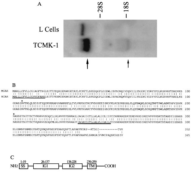
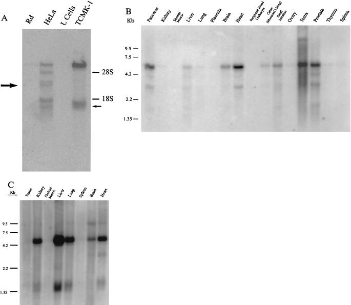
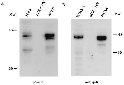
The subgroup C of the adenoviruses (Ad) and the group B coxsackieviruses (CVB) are structurally unrelated viruses that are known to compete for an unidentified cell surface receptor. We now describe the isolation of cDNAs from human and mouse that encode the human CVB and Ad2 and 5 receptor (HCAR) and the mouse CVB Ad2 and 5 receptor (MCAR). Both are 46-kDa glycoproteins whose primary amino acid sequences are highly homologous. Structurally, HCAR and MCAR appear to be transmembrane proteins that contain two extracellular immunoglobulin-like domains and therefore belong to this superfamily. Transfection of either of these cDNA molecules into receptor-negative NIH 3T3 cells conferred susceptibility to CVB infection and permitted the expression of beta-galactosidase from a recombinant Ad5 vector. In addition, HCAR and MCAR mRNAs could be detected on Northern blots of oligo(dT)-selected RNA from receptor-positive HeLa cells and TCMK-1 as well as several tissues of human and mouse origin that are known to be targets for Ad and CVB infections. Finally, Western blots using antibodies that inhibit virus binding to either the human or mouse CVB receptors detected 46-kDa proteins in HCAR- and MCAR-transfected cells, respectively. Taken together, these results confirm that the isolated cDNAs encode the receptors for the subgroup C Ad and CVB.


Figures

Figure 1
Figure 1
(A) Unique RNAs are found in receptor-expressing TCMK-1 cells. Total RNA from TCMK-1 or mouse L cells was probed with a 32P-labeled fragment of RTMCR-4 to reveal differentially expressed 6-kb (large arrow) and 1.4-kb (small arrow) RNAs. (B) Amino acid alignment of HCAR and MCAR. The deduced amino acid sequences of the cloned HCAR and MCAR cDNAs were aligned using the GeneWorks software program. Vertical lines, amino acid identities; dashes, gaps in the alignment. The positions of the potential signal peptide (thin underline), transmembrane region (thick underline), and N-linked glycosylation sites (∗) are shown. (C) A domain model for the CAR proteins. IG1 and IG2, immunoglobulin domains; SS, signal sequence; TM, transmembrane-spanning region.
Figure 4
Figure 4
HCAR and MCAR mRNA is expressed in a variety of tissues. (A) Hybridizing mRNAs of 6 kb and 1.4 kb can be detected in poly(A)+ Northern blots of TCMK-1 cells, but not mouse L cells. In contrast, six species of mRNAs are detected in HeLa cells that range in size from 6 kb to 1.2 kb that are not found in receptor-negative rhabdomyosarcoma (Rd) cells. The sizes of the RNAs that correlate with the size of the isolated HCAR (large arrow) and MCAR (small arrow) cDNAs are shown. (B) Human multiple tissue Northern blots were purchased from CLONTECH and hybridized with a 32P-labeled fragment that corresponded to the open reading frame of HCAR. Hybridizing sequences are similar in size to those detected in HeLa cells. (C) Mouse multiple tissue Northern blots (CLONTECH) were hybridized with a 32P-labeled fragment that corresponded to the open reading frame of MCAR. Hybridizing sequences are similar in size to those detected in TCMK-1 cells.
Figure 2
Figure 2
(A) pRTHR and pRTMR confer susceptibility to Ad5 entry. Only (A) TCMK-1 positive controls or NIH 3T3 cells transfected with either (B) pRTHR or (C) pRTMR stained for β-galactosidase expression after incubation with a CMV-β-galactosidase recombinant Ad5 vector at a multiplicity of infection of around 50. (D) pBK-CMV controls showed no reactivity after incubation under identical conditions. (B) HCAR expression is localized to the exterior of the cell. Bright staining was observed over the entire plasma membrane in unfixed NIH 3T3 cells transfected with pRTHR and labeling transfectants with RmcB and a fluorescein isothiocyanate-labeled goat anti-mouse antibody (A). B represents pBK-CMV control cells.
Figure 3
Figure 3
HCAR and MCAR are detectable by CVB receptor-specific antibodies. Forty-six-kilodalton proteins are visible in HCAR- and MCAR-transfected cells that are absent in pBK-CMV transfected cells. Similar sized proteins (≈46 kDa) are also detectable in HeLa cell and TCMK-1 cell positive controls. Immunodetection was performed using RmcB (A) (15) or anti-p46 (B) (6).

References

    1. Lonberg-Holm K, Crowell R L, Philipson L. Nature (London) 1976;259:679–681. - PubMed
    1. Xia D, Henry L J, Gerard R D, Deisenhofer J. Structure (London) 1994;2:1259–1270. - PubMed
    1. Muckelbauer J K, Kremer M, Minor I, Diana G, Dutko F J, Groarke J, Pevear D C, Rossman M G. Structure (London) 1995;3:653–667. - PubMed
    1. Shenk T. In: Virology. Fields B N, Knipe D M, Howley P M, Chanock R M, Melnick J L, Monath J P, Roizman B, Straus S E, editors. Philadelphia: Lippincott-Raven; 1996. pp. 2112–2137.
    1. Melnick J L. In: Virology. Fields B N, Knipe D M, Chanock R M, Hirsch M S, Melnick J L, Monath J P, editors. New York: Raven; 1990. pp. 549–600.

Associated data