. 1997 Apr 1;94(7):3363-7.
doi: 10.1073/pnas.94.7.3363.

Induction of dopamine D3 receptor expression as a mechanism of behavioral sensitization to levodopa

Affiliations

Induction of dopamine D3 receptor expression as a mechanism of behavioral sensitization to levodopa

R Bordet et al. Proc Natl Acad Sci U S A. .

Abstract

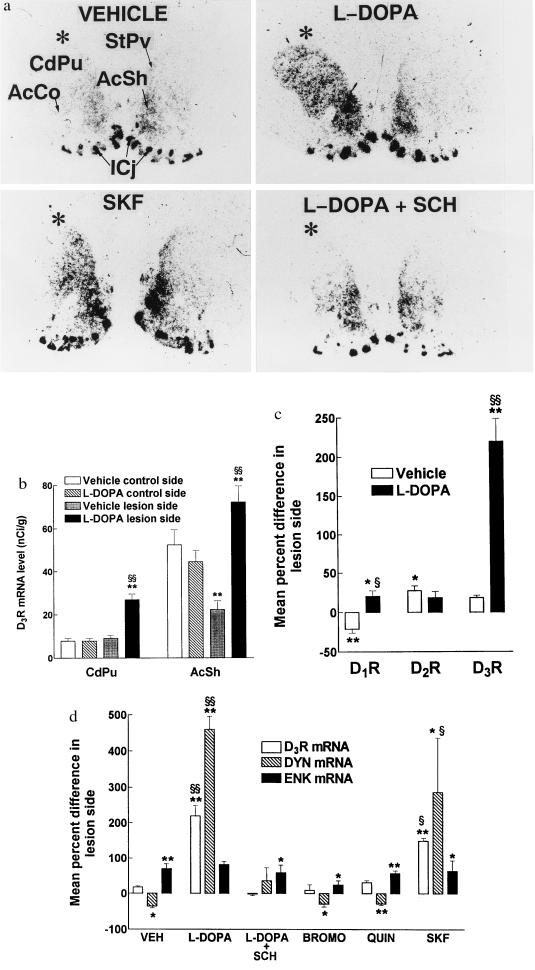
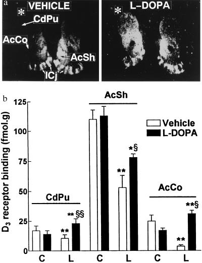
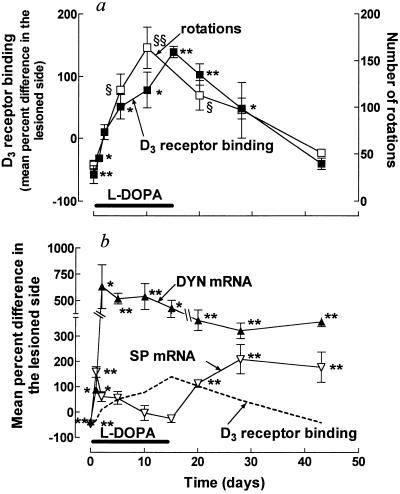
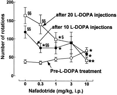
In rats with unilateral lesions of the nigrostriatal dopamine pathway with 6-hydroxydopamine, the motor stimulating effects of levodopa, an indirect dopamine receptor agonist, evidenced by contraversive rotations, become enhanced upon repeated intermittent administration. However, the mechanisms of this behavioral sensitization are essentially unknown. We show that development of sensitization is accompanied by a progressive appearance of D3 receptor mRNA and binding sites, visualized by in situ hybridization and 7-[3H] hydroxy-N,N-di-n-propyl-2-aminotetralin autoradiography, respectively, occurring in the denervated caudate putamen, a brain area from which this receptor subtype is normally absent. Development and decay of these two processes occur with closely parallel time courses, whereas there were no marked changes in D1 or D2 receptor mRNAs. D3 receptor induction by levodopa is mediated by repeated D1 receptor stimulation, since it is prevented by the antagonist SCH 33390 and mimicked by the agonist SKF 38393, but not by two D2 receptor agonists. The enhanced behavioral response to levodopa is mediated by the newly synthesized D3 receptor, since it is antagonized by nafadotride, a preferential D3 receptor antagonist, in low dosage, which has no such effect before D3 receptor induction. D3 receptor induction and behavioral sensitization are also accompanied by a sustained enhancement of prodynorphin mRNA level and a progressively decreasing expression of the preprotachykinin gene. We propose that imbalance between dynorphin and substance P release from the same striatonigral motor efferent pathway, related to D3 receptor induction, is responsible for behavioral sensitization.


Figures

Figure 1
Figure 1
In situ hybridization analysis of the effects of repeated administration of dopamine agonists on dopamine receptor and neuropeptide gene expression in the striatal complex of unilaterally 6-OHDA-lesioned rats. Coronal sections were hybridized with 33P-labeled cRNA probes for D1, D2, or D3 receptors (DRs), prodynorphin (DYN), or preproenkephalin (ENK) mRNAs, apposed to autoradiographic films and resulting pictures analyzed densitometrically. (a) Typical pictures showing the increased D3 receptor mRNA in the lesion side (indicated by asterisk) of the dorsolateral striatum (CdPu), StPv, and shell or core subdivisions of nucleus accumbens (AcSh, AcCo) after a 5-day levodopa (l-DOPA) treatment and the blockade of this effect by SCH 23390 (SCH). The effect of levodopa was partially reproduced by SKF 38393 (SKF). ICj, islands of Calleja. (b) Mean D3 receptor mRNA levels ± SEM (n = 4–6) in the two sides of CdPu (excluding StPv) and AcSh following repeated administration of vehicle or l-DOPA. (c) Mean percent difference (±SEM) in D1, D2, and D3 receptor mRNA levels between lesion and control side of CdPu. Same animals as in b. The mean (±SEM) signals on the control side of vehicle-treated rats corresponded to 454 ± 54, 597 ± 63, and 8 ± 1 nCi/g (1 Ci = 37 GBq) for D1R, D2R, and D3R, respectively. (d) Mean percent difference (± SEM, n = 4–6) in D3R, DYN, and ENK mRNA levels between lesion and control side of CdPu following 5-day administration of levodopa alone or together with SCH of bromocriptine (BROMO), quinelorane (QUIN), or SKF. The mean (± SEM) in situ hybridization signal on the control side of vehicle-treated rats corresponded to 0.22 ± 0.03 and 1.7 ± 0.2 μCi/g for DYN and ENK, respectively. ∗, P < 0.05; ∗∗, P < 0.01 in lesion vs. control side by the paired Student’s t test; §, P < 0.05; §§, P < 0.01 in lesion side of drug vs. vehicle-treated animals by the Mann–Whitney U test.
Figure 2
Figure 2
Changes in D3 receptor binding elicited by repeated levodopa treatments of 6-OHDA-lesioned rats. (a) Digitized autoradiographic D3 receptor binding picture obtained with [3H]7-OH-DPAT, in animals treated twice a day for 5 days with vehicle (VEH) or levodopa (l-DOPA), in which nonspecific labeling has been subtracted. Asterisk indicates the lesion side. (b) Autoradiographic signals generated in a were quantified in CdPu, AcSh, and AcCo of control (C) and lesion (L) sides. Mean ± SEM of values from 4–6 animals. Nonspecific binding in CdPu was 50 and 15% of total in vehicle- and levodopa-treated animals, respectively. ∗, P < 0.05; ∗∗, P < 0.01 as compared with the control side by the paired Student’s t test; §, P < 0.05; §§, P < 0.01 in lesion side of drug vs. vehicle-treated animals by the Mann–Whitney U test.
Figure 3
Figure 3
Progressive changes in D3-receptor binding and levodopa-induced rotations and neuropeptide mRNAs in CdPu of unilaterally 6-OHDA-lesioned rats following repeated treatment with levodopa and withdrawal. Rats received levodopa (50 mg/kg i.p.) twice daily for up to 15 days (horizontal bar) and were challenged with a single same dose of levodopa at the indicated time during this chronic treatment or after withdrawal. Animals were killed 4 h after the challenge, D3 receptor autoradiography using [3H]7-OH-DPAT or in situ hybridization with probes for prodynorphin (DYN) or preprotachykinin (SP) mRNAs. Note that rotations were not measured in all groups of animals. No significant changes were observed in D3 receptor binding or in situ hybridization signals in the control side. Means ± SEM of values from 4–9 animals. ∗, P < 0.05; ∗∗, P < 0.01 as compared with the control side by the paired Student’s t test; §, P < 0.05; §§, P < 0.01 compared with values obtained following the first levodopa injection by the Mann–Whitney U test.
Figure 4
Figure 4
Effects of nafadotride, a preferential D3-receptor antagonist on levodopa-induced rotations. Contralateral rotations were measured in animals challenged with levodopa (50 mg/kg i.p.), alone or in combination with nafadotride at indicated doses on three occasions: the first, 4 weeks after the lesion (pre l-DOPA treatment); the second and third, after subsequent treatments with 10 or 20 levodopa (l-DOPA) injections (50 mg/kg i.p., twice daily). Means ± SEM of values from 4–6 animals. ∗, P < 0.05; ∗∗, P < 0.01 nafadotride vs. saline-treated animals; §, P < 0.05; §§, P < 0.01 in levodopa-treated vs. untreated animals by the Mann–Whitney U test.

References

    1. Robinson T E, Becker J B. Brain Res Rev. 1986;11:157–198. - PubMed
    1. Kalivas P W, Stewart J. Brain Res Rev. 1991;16:223–244. - PubMed
    1. Hornykiewicz O. Pharmacol Rev. 1966;18:925–964. - PubMed
    1. Hardie R J. In: Parkinson’s Disease. Stern G M, editor. Baltimore: The Johns Hopkins Univ. Press; 1990. pp. 559–596.
    1. Gancher S T. In: Parkinson’s Disease Neurobehavioral Aspects. Huber S T, Cummings J L, editors. New York: Oxford Univ. Press; 1992. pp. 273–287.

Publication types

LinkOut - more resources