. 1997 Apr 1;94(7):3396-401.
doi: 10.1073/pnas.94.7.3396.

Neuronal nitric oxide synthase alternatively spliced forms: prominent functional localizations in the brain

Affiliations

Neuronal nitric oxide synthase alternatively spliced forms: prominent functional localizations in the brain

M J Eliasson et al. Proc Natl Acad Sci U S A. .

Abstract

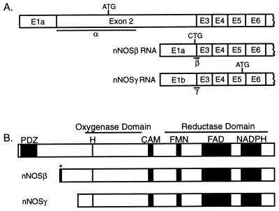
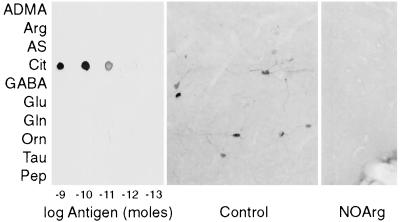
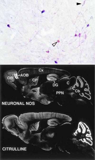
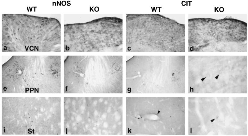
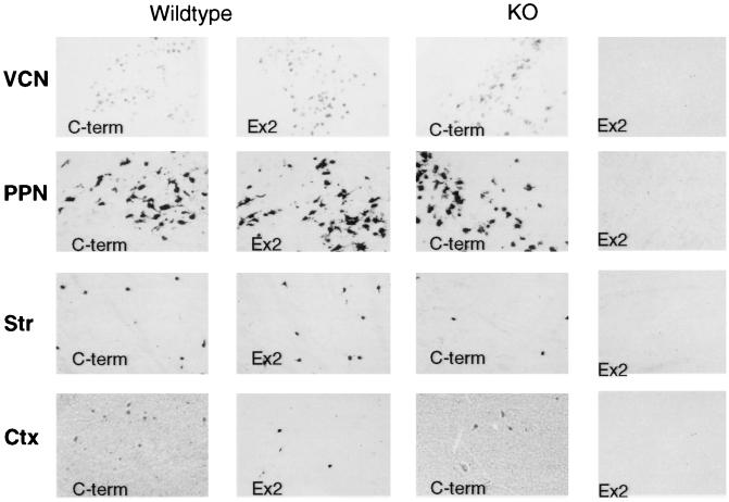
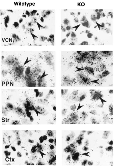
Neuronal nitric-oxide synthase (nNOS) is subject to alternative splicing. In mice with targeted deletions of exon 2 (nNOS(delta/delta)), two alternatively spliced forms, nNOS beta and gamma, which lack exon 2, have been described. We have compared localizations of native nNOS alpha and nNOS beta and gamma by in situ hybridization and immunohistochemistry in wild-type and nNOS(delta/delta) mice. To assess nNOS catalytic activity in intact animals we localized citrulline, which is formed stoichiometrically with NO, by immunohistochemistry. nNOS beta is prominent in several brain regions of wild-type animals and shows 2-to 3-fold up-regulation in the cortex and striatum of nNOS(delta/delta) animals. The persistence of much nNOS mRNA and protein, and distinct citrulline immunoreactivity (cit-IR) in the ventral cochlear nuclei and some cit-IR in the striatum and lateral tegmental nuclei, indicate that nNOS beta is a major functional form of the enzyme in these regions. Thus, nNOS beta, and possibly other uncharacterized splice forms, appear to be important physiological sources of NO in discrete brain regions and may account for the relatively modest level of impairment in nNOS(delta/delta) animals.


Figures

Figure 1
Figure 1
Isoforms of neuronal NOS. (A) Illustration of mRNAs generated by alternative splicing of the nNOS gene. (Top) The predominant form in wt. Location of start codons and regions recognized by the nNOS α, β, and γ in situ probes are indicated. (Adapted from refs. and .) (B) Illustration of nNOS isoforms. Consensus binding sites for heme (H), calmodulin (CAM), flavin mononucleotide (FMN), flavin adenine dinucleotide (FAD), and the reduced form of NADPH are indicated. nNOSβγ both lack the PDZ domain (PDZ), but the primary structure of the catalytic domains appears intact. ∗, six amino acids unique to nNOSβ. Based on refs. –.
Figure 2
Figure 2
Citrulline antiserum specificity. (Left) Dialyzed rat brain cytosol was coupled to various ligands with glutaraldehyde and then reduced with NaBH4. Serial 1:10 dilutions of each conjugate were spotted onto three pieces of nitrocellulose and then probed with a 1:10,000 dilution of antiserum. ADMA, asymmetric NG,NG-dimethylarginine; Arg, arginine; AS, argininosuccinate; Cit, citrulline; GABA, γ-aminobutyrate; Glu, glutamate; Gln, glutamine; Orn, ornithine; Tau, taurine; Pep, human glycine decarboxylase (residues 1,000–1,020). (Center) Mouse basal forebrain/islands of Calleja stained with 1:10,000 dilution of antiserum. Neuronal soma and processes are clearly labeled. (Right) Same area in mouse treated with nitroarginine, 50 mg/kg i.p. twice a day for 4 days. No neuronal staining is evident in this or other areas (data not shown) of the brain.
Figure 3
Figure 3
Comparison of cit-IR and nNOS-IR. (Top) Double labeling for nNOS (purple) and citrulline (light brown-yellow). All cit-IR cells observed were also nNOS-IR, although many nNOS-IR cells devoid of cit-IR were observed (solid arrowhead). Cit-IR was especially high in the soma (open arrowhead), while nNOS-IR appeared both in the soma and processes. (Bottom) nNOS-IR and cit-IR in sagital sections of adult mouse. White areas represent positive staining. OB, olfactory bulb; AOB, accessory olfactory bulb; CP, caudate-putamen; DG, dentate gyrus; Cx, cerebral cortex; C, colliculi; PPN, pedunculopontine nuclei; Cb, cerebellum.
Figure 4
Figure 4
Comparison of cit-IR and nNOS-IR in wild-type (WT) and nNOSΔ/Δ (KO) brains. VCN, ventral cochlear nucleus; PPN, pedunculopontine and laterodorsal tegmental nuclei; St, striatum. (h and l) High magnifications of the laterodorsal tegmental nucleus and striatum, respectively. Arrowheads (h and l) indicate cit-IR soma in nNOSΔ/Δ. Arrowhead (k) indicates one long, continuous cit-IR process. Dark areas indicate positive staining.
Figure 5
Figure 5
Comparison of mRNA localizations for nNOSa and total nNOS. cRNA digoxygenin in situ hybridization employed probes directed against nNOS C-terminal (C-term), representing total nNOS mRNA, and an nNOSα-specific probe (ex2) in wt and nNOSΔ/Δ (KO) animals. VCN, ventral cochlear nucleus; PPN, pedunculopontine nuclei; Str, striatum; Ctx, cerebral cortex. (×100.)
Figure 6
Figure 6
nNOSβ mRNA localizations in wild-type and nNOSΔ/Δ (KO) mice. Radioactive oligonucleotide in situ hybridization employed the nNOSβ-specific probe. Arrowheads indicate selected cells showing black radioactive emulsion grains. Sections are counterstained with cresyl violet. Abbreviations are as in Fig. 5 legend. (×400.)

References

    1. Bredt D S, Snyder S H. Proc Natl Acad Sci USA. 1990;87:682–685. - PMC - PubMed
    1. Schmidt H H H W, Walter U. Cell. 1994;78:919–925. - PubMed
    1. Garthwaite J, Boulton C L. Annu Rev Physiol. 1995;57:683–706. - PubMed
    1. Brenman J E, Chao D S, Gee S H, McGee A W, Craven S E, Santillano D R, Wu Z, Huang F, Xia H, Peters M F, Froehner S C, Bredt D S. Cell. 1996;84:757–767. - PubMed
    1. Silvagno F, Xia H, Bredt D S. J Biol Chem. 1996;271:11204–11208. - PubMed

Publication types

LinkOut - more resources