. 1997 Apr 1;94(7):3425-30.
doi: 10.1073/pnas.94.7.3425.

Mammary-derived signals activate programmed cell death during the first stage of mammary gland involution

Affiliations

Mammary-derived signals activate programmed cell death during the first stage of mammary gland involution

M Li et al. Proc Natl Acad Sci U S A. .

Abstract

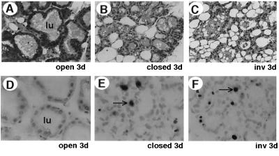
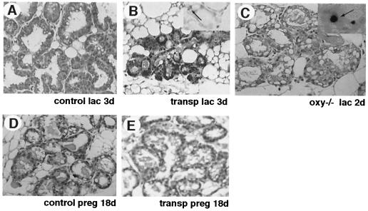
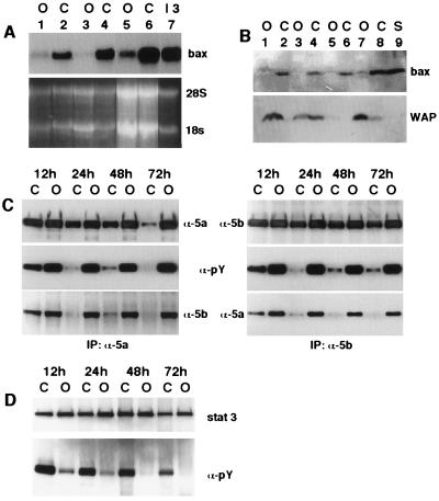
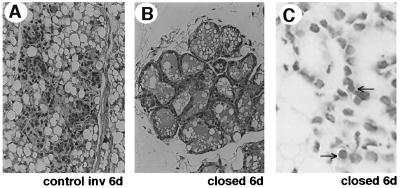
Programmed cell death (PCD) of mammary alveolar cells during involution commences within hours of the end of suckling. Locally, milk accumulates within alveolar lumens; systemically, levels of lactogenic hormones fall. Four experimental models were used to define the role of local factors as compared with systemic hormones during the first and second stages of involution. In three models, milk release was disrupted in the presence of systemic lactogenic hormones: (i) sealing of the teats, (ii) mammary gland transplants that cannot release milk due to the absence of a teat connection, and (iii) inactivation of the oxytocin gene. The ability of systemic hormones to preserve lobular-alveolar structure without blocking PCD was illustrated using a fourth transgenic model of lactation failure. During the first stage of involution, local signals were sufficient to induce alveolar PCD even in the presence of systemic lactogenic hormones. PCD coincided with bax induction, decreased expression of milk proteins, block of prolactin signal transduction through Stat5a and 5b, and activation of Stat3. The two stages of mammary gland involution are regulated by progressive gain of death signals and loss of survival factors. This study demonstrates that genetic events that occur during the first reversible stage are controlled by local factors. These mammary-derived death signals are dominant over protective effects related to systemic hormone stimulation.


Figures

Figure 1
Figure 1
Mammary-derived signals activate PCD during the first stage of involution. Histological analysis of mammary tissue from 3 days open (A and D) and closed glands (B and E) compared with normal 3 days involution (C and F). Hematoxylin and eosin staining (A, B, and C). In situ detection of PCD (D, E, and F). Arrows indicate cells undergoing PCD. Open, open teat; closed, Nexaband sealed teat; inv, normal involution after pup removal; 3d, 3 days; Lu, lumen.
Figure 2
Figure 2
Systemic hormone stimulation does not block alveolar PCD. Histological analysis of mammary tissue from 3 days postparturition transplanted (B) and nonmanipulated control (A) glands and 2 days postparturition oxytocin-deficient (oxy −/−) gland (C). Mammary gland development during pregnancy is normal in transplanted glands (E) as compared with control (D). Hematoxylin and eosin staining (A-E). In situ detection of PCD (Insets in B and C). Arrows indicate cells undergoing PCD. Control, nonmanipulated gland in a mouse receiving a transplant; lac 3d (2d), 3 (2) days postpartum lactation; transp, transplanted gland; preg 18d, 18 days pregnancy.
Figure 3
Figure 3
Local stimuli are sufficient for induction of bax gene expression and phosphorylation changes in Stat5a/5b and Stat3. (A) Northern blot analysis of steady state levels of bax mRNA in open (O), closed (C), and normal involution day 3 (I3) glands. Loading control, ethidium bromide staining of 18S and 28S RNA. (B) Western blot analysis of steady state levels of bax and WAP protein in open (O) and closed (C) glands compared with spleen (S). Coomassie blue staining of the protein gel demonstrated equal loading of all samples. (C) Analysis of Stat5a and b protein levels, phosphorylation, and heterodimerization by immunoprecipitation and Western blot in open (O) and closed (C) glands at 12, 24, 48, and 72 h. α-5a, anti-Stat5a Ab; α-5b, anti-Stat5b Ab; α-pY, antiphosphotyrosine Ab. (D) Analysis of Stat3 protein levels and phosphorylation by immunoprecipitation and Western blot in open (O) and closed (C) glands at 12, 24, 48, and 72 h. α-3: anti-Stat3 Ab.
Figure 4
Figure 4
Maintenance of lactogenic hormone stimulation blocks progression into the second stage of involution. Histological analysis of mammary tissue from 6 days normal involution (A) and 6 days closed glands with maintenance of suckling (B and C). Hematoxylin and eosin staining (A and B). In situ detection of PCD (C). Arrows indicate cells undergoing PCD. Control inv 6d, normal 6 days involution; closed 6 d, 6 days post teat closure.
Figure 5
Figure 5
Systemic hormonal stimulation by suckling or glucocorticoids alone preserve lobular-alveolar structure without blocking PCD. Histological analyses of mammary tissue from WAP-TAg transgenic mice in the absence (A) and presence (B and C) of suckling or after exogenous glucocorticoids (D). Hematoxylin and eosin staining (A and B). In situ detection of PCD (C and D). Arrows indicate cells undergoing PCD. TAg, WAP-TAg transgenic mouse; inv 3d, 3 days involution; suc 3d, 3 days suckling; +H, hydrocortisone injected mouse. (E) Northern blot analyses of β-casein expression in 3 days suckled TAg mice. Loading control, ethidium bromide staining of 18S and 28S RNA; P, day 18 pregnancy; suckling 3d, samples from individual TAg mice suckled for 3 days; I 3d, 3 days involution.
Figure 6
Figure 6
Extended alveolar cell survival through continuous suckling. Hematoxylin- and eosin-stained sections of mammary tissue from 25 days postpartum mouse with a single litter (A) and 49 days postpartum mouse with serial litter replacement (B). lac 25d, suckling by a single litter for 25 days; lac 49d, suckling from multiple litters for 49 days. (C) Model of mammary gland involution illustrating progression through the first and second stages.

References

    1. Feng Z, Marti A, Jehn B, Altermatt H J, Chicaiza G, Jaggi R. J Cell Biol. 1995;131:1095–1103. - PMC - PubMed
    1. Lund L R, Romer J, Dohy-Thomasset N, Solberg H, Pyke C, Bissell M J, Dane K, Werb Z. Development. 1996;122:181–193. - PMC - PubMed
    1. Strange R, Li F, Saurer S, Burkhardt A, Friis R. Development. 1992;115:49–58. - PubMed
    1. Marti A, Jehn B, Costello E, Keon N, Ke G, Martin F, Jaggi R. Oncogene. 1994;9:1213–1223. - PubMed
    1. Marti A, Feng Z, Jehn B, Djonov V, Chicaiza G, Altermatt H J, Jaggi R. Cell Death Differ. 1995;2:277–283. - PubMed

Publication types