. 1997 Apr 7;185(7):1231-9.
doi: 10.1084/jem.185.7.1231.

Intracellular antimicrobial activity in the absence of interferon-gamma: effect of interleukin-12 in experimental visceral leishmaniasis in interferon-gamma gene-disrupted mice

Affiliations

Intracellular antimicrobial activity in the absence of interferon-gamma: effect of interleukin-12 in experimental visceral leishmaniasis in interferon-gamma gene-disrupted mice

A P Taylor et al. J Exp Med. .

Abstract

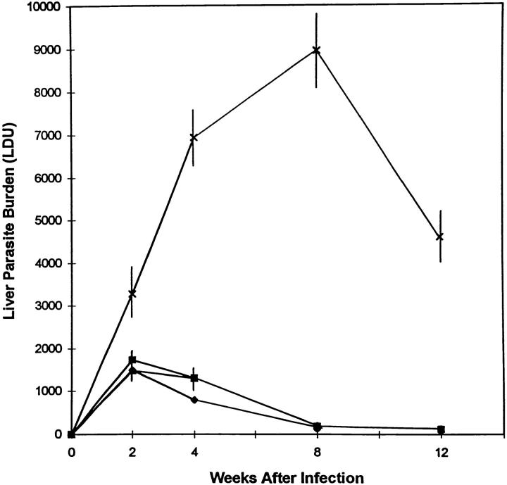
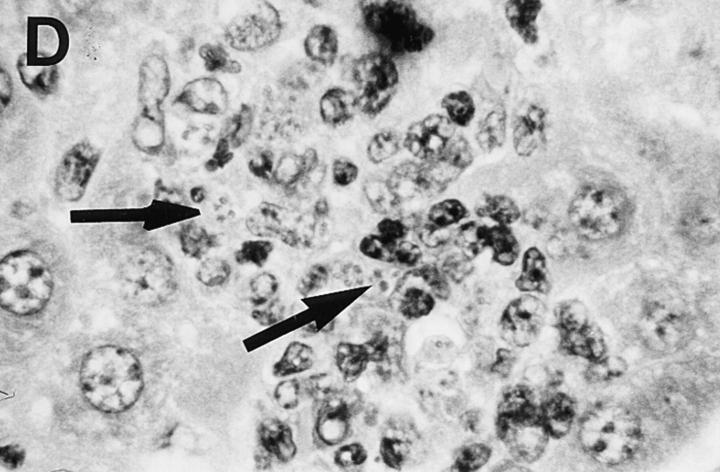
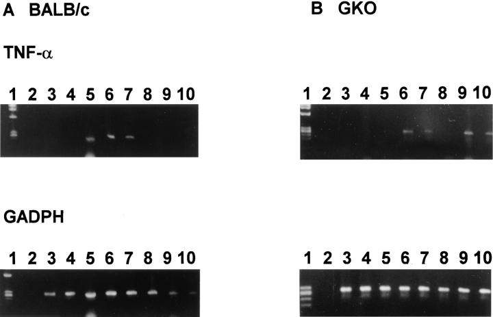
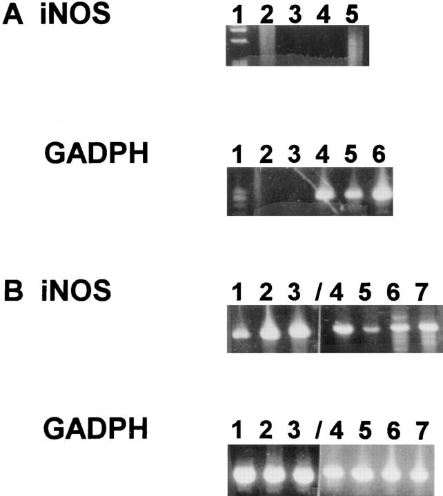
Despite permitting uncontrolled intracellular visceral infection for 8 wk, interferon-gamma (IFN-gamma) gene knockout (GKO) mice infected with Leishmania donovani proceeded to reduce liver parasite burdens by 50% by week 12. This late-developing IFN-gamma-independent antileishmanial mechanism appeared to be dependent largely on endogenous tumor necrosis factor-alpha (TNF-alpha): L. donovani infection induced TNF-alpha mRNA expression in parasitized GKO livers and neutralization of TNF-alpha reversed control at week 12.7 d of treatment of infected GKO mice with interleukin-12 (IL-12) readily induced leishmanicidal activity and also partially restored the near-absent tissue granulomatous response, observations that for the first time expand the antimicrobial repertoire of IL-12 to include IFN-gamma-independent effects. The action of IL-12 against L. donovani was TNF-alpha dependent and required the activity of inducible nitric oxide synthase. These results point to the presence of an IFN-gamma-independent antimicrobial mechanism, mediated by TNF-alpha, which remains quiescent until activated late in the course of experimental visceral leishmaniasis. However, as judged by the effect of exogenous IL-12 this quiescent mechanism can readily be induced to rapidly yield enhanced intracellular antimicrobial activity.


Figures

Figure 1
Figure 1
Course of visceral infection in GKO mice. Results represent mean LDU ± SEM from 6–12 animals per timepoint for GKO−/− (X), GKO+/+ (▪), and BALB/c mice (♦); except at week 12 for BALB/c mice (n = 4) and GKO+/− mice (▴, identical to GKO+/+ or BALB/c LDU at all timepoints) (n = 3).
Figure 2
Figure 2
Liver histologic reaction in L. donovani–infected GKO mice and response to IL-12 treatment. (A) Normal BALB/c mouse shows mature granuloma formation surrounding fused parasitized Kupffer cell core 4 wk after infection. (B) 4-wk–infected GKO liver showing three large heavily parasitized foci (arrows) with minimal mononuclear cell infiltrate. (C) 3-wk–infected untreated GKO mouse showing heavily infected Kupffer cell focus with no cellular infiltrate. (D) 3-wk–infected GKO mouse treated with IL-12 during the preceding 7 d shows few parasites (arrow) and clearly enhanced cell influx. Original magnification, ×630.
Figure 2
Figure 2
Liver histologic reaction in L. donovani–infected GKO mice and response to IL-12 treatment. (A) Normal BALB/c mouse shows mature granuloma formation surrounding fused parasitized Kupffer cell core 4 wk after infection. (B) 4-wk–infected GKO liver showing three large heavily parasitized foci (arrows) with minimal mononuclear cell infiltrate. (C) 3-wk–infected untreated GKO mouse showing heavily infected Kupffer cell focus with no cellular infiltrate. (D) 3-wk–infected GKO mouse treated with IL-12 during the preceding 7 d shows few parasites (arrow) and clearly enhanced cell influx. Original magnification, ×630.
Figure 3
Figure 3
Kinetics of TNF-α mRNA expression in BALB/c mice (A) and GKO mice (B). Qualitative RT-PCR was performed using cDNA from liver RNA extracted at each timepoint. Each lane represents one mouse. Lane 1, size markers; lane 2, PCR control; lanes 3 and 4, 2 wk after infection; lanes 5 and 6, 4 wk after infection; lanes 7 and 8, 8 wk after infection; lanes 9 and 10, 12 wk after infection. Similar results were obtained from six mice for each timepoint from 2–3 separate experiments.
Figure 4
Figure 4
Expression of iNOS mRNA in livers of L. donovani–infected BALB/c mice. Total liver RNA was isolated at 2, 3, and 4 wk after infection and assayed for the presence of iNOS message by qualitative RTPCR. Each lane represents one mouse. GADPH is a control for the amount of cDNA in each sample. (A) iNOS RT-PCR: lane 1, size markers; lane 2, PCR control; lanes 3, 4, and 5, 2-wk samples. GADPH RTPCR: lane 1, size markers; lane 2, PCR control; lane 3, blank, lanes 4, 5, and 6, 2-wk samples. (B) iNOS RT-PCR: lanes 1, 2, and 3: 3-wk samples; lanes 4, 5, 6, and 7, 4-wk samples from a different gel and PCR reaction. GADPH RT-PCR, same as iNOS.

References

    1. Squires KE, Schreiber RD, McElrath MJ, Rubin BY, Anderson SL, Murray HW. Experimental visceral leishmaniasis: role of endogenous IFN-γ in host defense and tissue granulomatous response. J Immunol. 1989;143:4244–4249. - PubMed
    1. Murray HW. Effect of continuous administration of interferon-γ in experimental visceral leishmaniasis. J Infect Dis. 1990;161:992–994. - PubMed
    1. Dalton DK, Pitts-Meek S, Keshav S, Figari IS, Bradley A, Stewart TA. Multiple defects of immune cell function in mice with disrupted interferon-γ genes. Science (Wash DC) 1993;159:1739–1742. - PubMed
    1. Graham MB, Dalton DK, Giltinan D, Braciale VL, Stewart TA, Braciale TJ. Response to influenza infection in mice with a targeted disruption in the interferon-γ gene. J Exp Med. 1993;178:1725–1732. - PMC - PubMed
    1. Cooper. A.M., D.K. Dalton, T.A. Stewart, J.P. Griffin, D.G. Russell, and I.M. Orme. Disseminated tuberculosis in interferon-γ gene-disrupted mice. J Exp Med. 1993;178:2243–2247. - PMC - PubMed

Publication types

MeSH terms