Comparative Study
. 1997 Apr 7;185(7):1295-305.
doi: 10.1084/jem.185.7.1295.

The human immunodeficiency virus type 1 (HIV-1) Vpu protein interferes with an early step in the biosynthesis of major histocompatibility complex (MHC) class I molecules

Affiliations
Comparative Study

The human immunodeficiency virus type 1 (HIV-1) Vpu protein interferes with an early step in the biosynthesis of major histocompatibility complex (MHC) class I molecules

T Kerkau et al. J Exp Med. .

Abstract

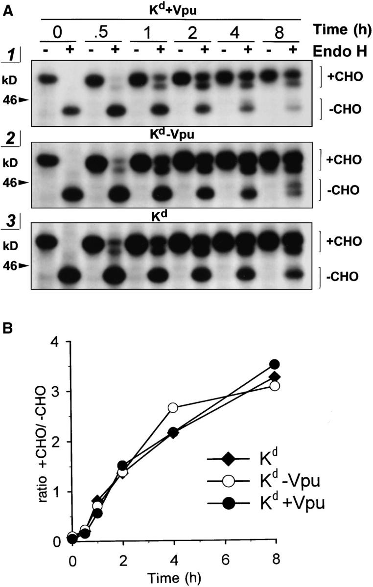
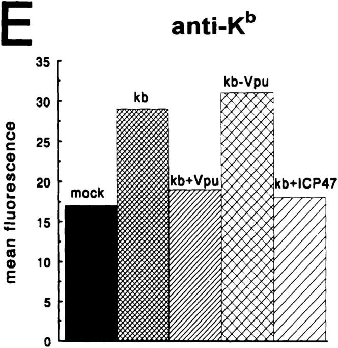
The human immunodeficiency virus type 1 (HIV-1) vpu gene encodes a small integral membrane phosphoprotein with two established functions: degradation of the viral coreceptor CD4 in the endoplasmic reticulum (ER) and augmentation of virus particle release from the plasma membrane of HIV-1-infected cells. We show here that Vpu is also largely responsible for the previously observed decrease in the expression of major histocompatibility complex (MHC) class I molecules on the surface of HIV-1-infected cells. Cells infected with HIV-1 isolates that fail to express Vpu, or that express genetically modified forms of Vpu that no longer induce CD4 degradation, exhibit little downregulation of MHC class I molecules. The effect of Vpu on class I biogenesis was analyzed in more detail using a Vpu-expressing recombinant vaccinia virus (VV). VV-expressed Vpu induces the rapid loss of newly synthesized endogenous or VV-expressed class I heavy chains in the ER, detectable either biochemically or by reduced cell surface expression. This effect is of similar rapidity and magnitude as the VV-expressed Vpu-induced degradation of CD4. Vpu had no discernible effects on cell surface expression of VV-expressed mouse CD54, demonstrating the selectivity of its effects on CD4 and class I heavy chains. VV-expressed Vpu does not detectably affect class I molecules that have been exported from the ER. The detrimental effects of Vpu on class I molecules could be distinguished from those caused by VV-expressed herpes virus protein ICP47, which acts by decreasing the supply of cytosolic peptides to class I molecules, indicating that Vpu functions in a distinct manner from ICP47. Based on these findings, we propose that Vpu-induced downregulation of class I molecules may be an important factor in the evolutionary selection of the HIV-1-specific vpu gene by contributing to the inability of CD8+ T cells to eradicate HIV-1 from infected individuals.


Figures

Figure 1
Figure 1
(A) Cell surface expression of MHC-I molecules is downregulated in TCD8+-depleted and PHA/IL-2–activated PBL infected with HIV-1, but not in PBL infected with HIV-2 or SIV. Uninfected control (a); PBL infected with HIV-2D205 (b); SIVmac (c); HIV-1BH10 (d); intracellular staining for viral core proteins p24/p27gag (e–h). The thin curves to the left in each histogram represent negative controls. (B) Downregulation of MHC-I expression on HIV-1 PBL depends on the expression of biologically active Vpu protein. Uninfected controls (a, e, and i); TCD8+depleted and PHA/IL-2–activated PBL infected with wild-type HIVNL4-3 (b, f, and j), the vpu mutant HIVNL-U35 (c, g, and k), and HIVNL4-3/U2/6 encoding nonphosphorylated Vpu2/6 (d, h, and l). The left curves in a, e, and i represent negative controls. Cells were stained with either conformation-specific mAb I0T2 (a–d). Virus expression was controlled by intracellular staining for p24gag (e–f).
Figure 2
Figure 2
Vpu compromises generation of MHC class I complexes in HIV-1 infected PBL. (A) Pulse/chase analysis of MHC-I molecules in TCD8+- depleted and PHA/IL-2–activated PBL infected with HIVNL4-3 (wild type, +Vpu), or the mutant HIVNL-U35 (−Vpu); uninfected control (mock). MHC class I complexes were immunoprecipitated with mAb W6/32, separated in a 10% acryl aide gel, and analyzed by fluorography. Only parts of the fluorograms demonstrating H chain–specific bands in the range of 43 kD are shown. (B) Relative amounts of H chain proteins seen in (A) were quantitated by means of an image analyzer and blotted against the time of the chase period (pu, nonstandardized arbitrary PhosphoImager units). (C) TCD8+depleted and PHA/IL-2–activated PBL isolated from another donor were infected with HIVNL4-3 (+Vpu), HIVNL-U35 (−Vpu), or HIVNL4-3/U2/6 expressing a nonphosphorylated mutant (+Vpu2/6). Pulse/chase experiment, immunocollection, and SDS-PAGE analysis (not shown) were performed as described for Fig. 2, A and B. Relative amounts of H chains were quantitated with a PhosphorImager and plotted in arbitrary units (pu) against the time of the chase period.
Figure 2
Figure 2
Vpu compromises generation of MHC class I complexes in HIV-1 infected PBL. (A) Pulse/chase analysis of MHC-I molecules in TCD8+- depleted and PHA/IL-2–activated PBL infected with HIVNL4-3 (wild type, +Vpu), or the mutant HIVNL-U35 (−Vpu); uninfected control (mock). MHC class I complexes were immunoprecipitated with mAb W6/32, separated in a 10% acryl aide gel, and analyzed by fluorography. Only parts of the fluorograms demonstrating H chain–specific bands in the range of 43 kD are shown. (B) Relative amounts of H chain proteins seen in (A) were quantitated by means of an image analyzer and blotted against the time of the chase period (pu, nonstandardized arbitrary PhosphoImager units). (C) TCD8+depleted and PHA/IL-2–activated PBL isolated from another donor were infected with HIVNL4-3 (+Vpu), HIVNL-U35 (−Vpu), or HIVNL4-3/U2/6 expressing a nonphosphorylated mutant (+Vpu2/6). Pulse/chase experiment, immunocollection, and SDS-PAGE analysis (not shown) were performed as described for Fig. 2, A and B. Relative amounts of H chains were quantitated with a PhosphorImager and plotted in arbitrary units (pu) against the time of the chase period.
Figure 3
Figure 3
(A) Expression of Vpu by recombinant vaccinia viruses. HeLa cells were infected with rVVs expressing either wild-type Vpu (VV-Vpu) or the mutant VpuDEL1 (VV-UDEL1). 3 h after infection, HeLa cells were pulse labeled with [35S]methionine for 25 min and cells expressing wild-type Vpu were chased for up to 3 h. Cell lysates were immunoprecipitated with antiVpu sera (sheep and rabbit), separated in a 12.5% acryl aide gel, and analyzed by fluorography. Positions of 14C-labeled molecular weight marker proteins are indicated on the left (M). (B) Mouse L929 cells were co- infected with rVVs expressing either human CD4 (vCB-3) or wild-type Vpu (VV-Vpu). 3 h after infection, cells were pulse labeled with [35S]methionine for 7 min and chased for up to 4 h. CD4 molecules were recovered with anti-CD4 serum, separated in a 10% acryl aide gel, and analyzed by fluorography. Only a part of the fluorogram demonstrating CD4-specific bands in the range of 55 kD is shown. Stability of CD4 present at different times during the chase period were calculated relative to the levels of CD4 present at the end of the pulse labeling (0), which was empirically defined as 100%.
Figure 3
Figure 3
(A) Expression of Vpu by recombinant vaccinia viruses. HeLa cells were infected with rVVs expressing either wild-type Vpu (VV-Vpu) or the mutant VpuDEL1 (VV-UDEL1). 3 h after infection, HeLa cells were pulse labeled with [35S]methionine for 25 min and cells expressing wild-type Vpu were chased for up to 3 h. Cell lysates were immunoprecipitated with antiVpu sera (sheep and rabbit), separated in a 12.5% acryl aide gel, and analyzed by fluorography. Positions of 14C-labeled molecular weight marker proteins are indicated on the left (M). (B) Mouse L929 cells were co- infected with rVVs expressing either human CD4 (vCB-3) or wild-type Vpu (VV-Vpu). 3 h after infection, cells were pulse labeled with [35S]methionine for 7 min and chased for up to 4 h. CD4 molecules were recovered with anti-CD4 serum, separated in a 10% acryl aide gel, and analyzed by fluorography. Only a part of the fluorogram demonstrating CD4-specific bands in the range of 55 kD is shown. Stability of CD4 present at different times during the chase period were calculated relative to the levels of CD4 present at the end of the pulse labeling (0), which was empirically defined as 100%.
Figure 4
Figure 4
Vpu induces loss of MHC-I H chains in HeLa cells. Parallel cultures of HeLa cells were infected with rVVs VV-Vpu (+Vpu) or VVUDEL1 (−Vpu), and 2.5 h after infection cells were pulse labeled with [35S]methionine for 10 min and chased for up to 8 h. Aliquots of cell lysates were immunoprecipitated with mAb W6/32 (A), polyclonal antibodies specific for human β2m (B), and mAb SK81075 specific for human H chains (C), separated in a 10% acryl aide gel, and analyzed by fluorography. Only bands corresponding to H chains in the range of 43 kD are demonstrated in the upper part; the quantitation of H chains by means of a PhosphorImager is demonstrated in the lower part. (D) Cell lysates were immunoprecipitated with antibodies specific for Vpu and the mAb TW2.3 specific for vaccinia structural proteins E3L and E3L*. Proteins were separated in a 12.5% acryl aide gel. Parts of the fluorogram depicting a range between 14 and 30 kD are shown in the upper part; the quantitation of the VV E3L is shown in the lower part.
Figure 5
Figure 5
Vpu disturbs an early process in MHC-I biogenesis. Parallel cultures of HeLa cells were co-infected with VV-Kd expressing mouse H chain Kd together with VV-Vpu (+Vpu), VV-UDEL1 (−Vpu), or VVICP47 (+ICP47). 2.5 h after infection cells were pulse labeled with [35S]methionine for 4 min, aliquoted in ice-cold medium, and chased at 37°C for up to 2 h. Half of the cell lysates were immunoprecipitated with mAb 215 (A) or with anti-Kd serum pAb-ex8 (B). Two rounds of immunocollection were conducted and H chain molecules collected were analyzed by SDS-PAGE followed by fluorography. Only bands corresponding to H chains are demonstrated in the upper part; the quantitation of H chains detected after sequential collection (1st and 2nd) by means of a PhosphorImager is demonstrated in the lower part, left histograms. Stability of H chains recovered is demonstrated in the right histograms. Arrows indicate mature glycosylated H chains detected after 2 h of chase period.
Figure 6
Figure 6
Transport of class I H chains is not affected by Vpu. Parallel cultures of HeLa cells were co-infected with VV-Kd together with VVVpu (Kd+Vpu) or VV-UDEL1 (KdVpu), or VV-Kd alone (Kd). 2.5 h after infection, cells were pulse labeled with [35S]methionine for 15 min and chased for up to 8 h. Class I molecules were immunocollected with a 1:1 mixture of mAb 215 and anti-Kd serum pAb-ex8. Bead-bound material was split and either not treated (−) or treated with endo H (+). Samples were analyzed on a 8% acryl aide gel. Parts of the fluorograms depicting H chain molecules are shown in A. The four bands in the treated samples represent H chains partially or completely resistant (+CHO) or sensitive (−CHO) to endo H treatment. Relative amounts of H chains seen in A were quantitated and the quotient +CHO/−CHO was calculated and plotted against the time of the chase period (B).
Figure 7
Figure 7
Vpu downregulates cell surface expression of MHC-I molecules. HeLa cells were infected with rVVs expressing either wild-type Vpu (+Vpu), the vpu mutant VpuDEL1 (−Vpu), or the HSV protein ICP47 (+ICP47). In D and C cells were co-infected with rVV expressing the mouse glycoprotein ICAM-1 (CD54); in E cells were co-infected with rVV expressing mouse H chain Kb. Cells were stained 5 h after infection with antibodies specific for human β2m (A), human MHC-I (mAb W6/32; B), human transferin receptor (TFR; CD72; C), mouse H chain Kb (E), or mouse ICAM-1 (D).
Figure 7
Figure 7
Vpu downregulates cell surface expression of MHC-I molecules. HeLa cells were infected with rVVs expressing either wild-type Vpu (+Vpu), the vpu mutant VpuDEL1 (−Vpu), or the HSV protein ICP47 (+ICP47). In D and C cells were co-infected with rVV expressing the mouse glycoprotein ICAM-1 (CD54); in E cells were co-infected with rVV expressing mouse H chain Kb. Cells were stained 5 h after infection with antibodies specific for human β2m (A), human MHC-I (mAb W6/32; B), human transferin receptor (TFR; CD72; C), mouse H chain Kb (E), or mouse ICAM-1 (D).

Similar articles

Cited by

References

    1. Townsend A, Öhlén C, Bastin J, Ljunggren H-G, Foster L, Kärre K. Association of class I major histocompatibility heavy and light chains induced by viral peptides. Nature (Lond) 1989;340:443–448. - PubMed
    1. Yewdell JW, Bennink JR. Cell biology of antigen processing and presentation to MHC class I moleculerestricted T lymphocytes. Adv Immunol. 1992;52:1–23. - PubMed
    1. Heemels M-T, Ploegh H. Generation, translocation, and presentation of MHC class I–restricted peptides. Annu Rev Biochem. 1995;64:463–491. - PubMed
    1. Peters JM. Proteasomes: protein degradation machines of the cell. TIBS (Trends Biochem Sci) 1994;19:377–382. - PubMed
    1. Niedermann G, Butz S, Ihlenfeldt HG, Grimm R, Lucchiari M, Hoschützky H, Jung G, Maier B, Eichmann K. Contribution of proteasome-mediated proteolysis to the hierarchy of epitopes presented by major histocompatibility complex class I molecules. Immunity. 1995;2:289–299. - PubMed

Publication types

MeSH terms

Substances