. 1997 Apr 7;185(7):1307-16.
doi: 10.1084/jem.185.7.1307.

Diversification of T cell responses to carboxy-terminal determinants within the 65-kD heat-shock protein is involved in regulation of autoimmune arthritis

Affiliations

Diversification of T cell responses to carboxy-terminal determinants within the 65-kD heat-shock protein is involved in regulation of autoimmune arthritis

K D Moudgil et al. J Exp Med. .

Abstract

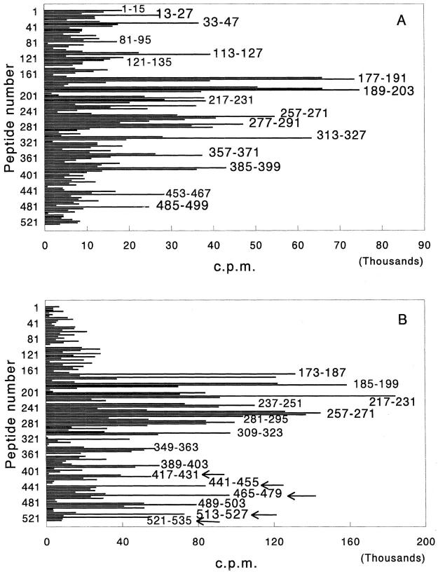
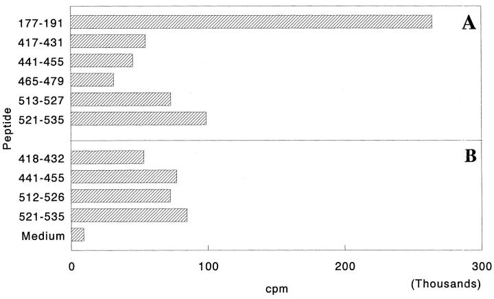
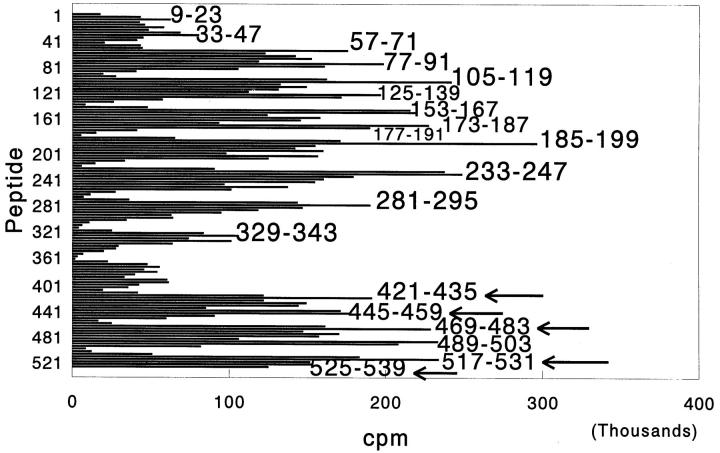
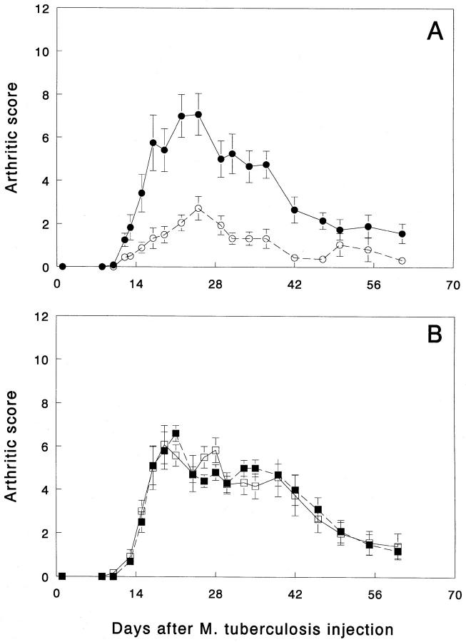
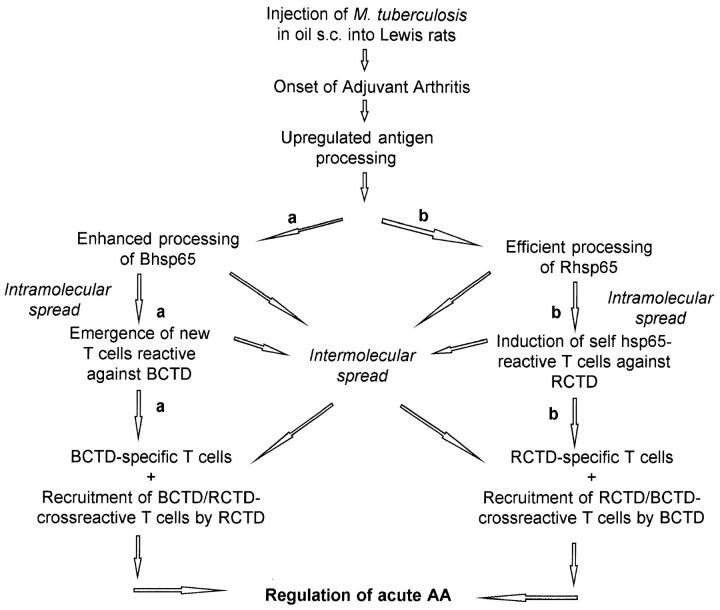
The T cell response to the 65-kD mycobacterial heat-shock protein (Bhsp65) has been implicated in the pathogenesis of autoimmune arthritis. Adjuvant arthritis (AA) induced in the Lewis rat (RT-1(l)) by injection of Mycobacterium tuberculosis serves as an experimental model for human rheumatoid arthritis (RA). However, the immunological basis of regulation of acute AA, or of susceptibility/resistance to AA is not known. We have defined the specificity of the proliferative T cell responses to Bhsp65 during the course of AA in the Lewis rat. During the early phase of the disease (6-9 d after onset of AA), Lewis rats raised T cell responses to many determinants within Bhsp65, spread throughout the molecule. Importantly, in the late phase of the disease (8-10 wk after onset of AA), there was evidence for diversification of the T cell responses toward Bhsp65 carboxy-terminal determinants (BCTD) (namely, 417-431, 441-455, 465-479, 513-527, and 521-535). Moreover, arthritic rats in the late phase of AA also raised vigorous T cell responses to those carboxy-terminal determinants within self(rat) hsp65 (Rhsp65) that correspond in position to the above BCTD. These results suggest that the observed diversification is possibly triggered in vivo by induction of self(Rhsp65)-reactive T cells. Interestingly, another strain of rat, the Wistar Kyoto (WKY/NHsd) rat (RT-1(l)), with the same major histocompatibility complex class II molecules as the Lewis rat, was found to be resistant to AA. In WKY rats, vigorous responses to the BCTD, to which the Lewis rat responded only in the late phase of AA, were observed very early, 10 d after injection of M. tuberculosis, Strikingly, pretreatment with the peptides comprising the set of BCTD, but not its amino-terminal determinants, provided significant protection to naive Lewis rats from subsequent induction of AA. Thus, T cell responses to the BCTD are involved in regulating inflammatory arthritis in the Lewis rat and in conferring resistance to AA in the WKY rat. These results have important implications in understanding the pathogenesis of RA and in devising new immunotherapeutic strategies for this disease.


Figures

Figure 1
Figure 1
Response of arthritic Lewis rats to Bhsp65 in the early (A) and late (B) phases of AA. Inbred Lewis rats were immunized subcutaneously with M. tuberculosis in a hind footpad, and then observed for signs of arthritis. 6–9 d (early phase; A) or 8–10 wk (late phase; B) after the appearance of clinical AA, rats were killed and their popliteal, inguinal, and axillary LNC pooled together were tested in a proliferation assay. In each group, LNC from four rats were pooled for testing with peptides. Overlapping pin peptides spanning the entire length of the Bhsp65 were used for the in vitro recall response in the assay. Each peptide is identified by its first amino acid residue. The results are expressed as cpm. The results of a representative experiment using pooled LNC from four rats are shown in each section of the figure. Similar results were obtained in repeat experiments with pooled LNC or upon testing cells from individual rats with selected Bhsp65 peptides (data not shown). In B, the T cell responses to new, unique Bhsp65 carboxy-terminal determinants (BCTD) are indicated by arrows.
Figure 2
Figure 2
Response of arthritic Lewis rats to peptides containing the COOH-terminal determinants within Bhsp65 (A) or Rhsp65 (B). Arthritis was induced as described in Materials and Methods. 4 wk after the appearance of clinical AA, rats were killed and their spleen cells (SPC) tested in a proliferation assay. The results are expressed as cpm. The results of a representative experiment are shown here. Similar results were obtained in repeat experiments (data not shown). The amino acid sequences of the five peptides comprising the BCTD and of the corresponding Rhsp65 peptides are given in Table 2. Response to peptide 177–191 is shown as a positive control. Rhsp65 peptide 465–479 could not be tested in this series of experiments.
Figure 3
Figure 3
Response of AA-resistant Wistar Kyoto (WKY) rats to Bhsp65 peptides after injection of M. tuberculosis. Rats were immunized with M. tuberculosis subcutaneously and after 10 d, the draining LNC from four rats were pooled and tested in a proliferation assay as described in Fig. 1. The results are expressed as cpm. The T cell responses to the unique carboxy-terminal determinants of Bhsp65 (to which arthritic Lewis rats respond only in the late phase of AA; shown in Fig. 1 B) are indicated by arrows. Although the highest proliferative responses correspond to the peptides marked by arrows, comparable or even higher responses also were raised to the adjacent overlapping peptides; namely, 417–431, 441– 455, 465–479, 513–527, and 521–535 in repeat experiments (data not shown). Responses to the BCTD were observed in repeat experiments in WKY rats tested 10–13 d after M. tuberculosis injection (data not shown).
Figure 4
Figure 4
Pretreatment of naive Lewis rats with peptides comprising the BCTD afford protection from subsequent induction of AA. One group of 5–6-wk-old Lewis rats (n = 9) was immunized subcutaneously with a combination of five Bhsp65 peptides (namely, 417–431, 441–455, 465–479, 513–527, and 521–535) (A) mixed in N,N-Dimethyl-N,Ndioctadecyl ammonium chloride (DDA) (GERBU adjuvant; GERBU Biotechnik GmbH, Gaiberg, Germany) (17, 55). Each rat received 100 μg of each of the five peptides mixed together in the same suspension. Another group of age- and sex-matched control Lewis rats (n = 6) was immunized with HEL peptide 85–96/DDA. After 5 wk, these rats were immunized subcutaneously with M. tuberculosis for induction of AA. From 7 d onwards, rats were examined daily or on alternate days for signs of arthritis. The severity of arthritis in each of the three uninjected paws was graded on a scale from 0 to 4 as described in Materials and Methods, and the highest score achievable in any rat was 12 (32, 33). Rats were observed for up to 61 d after injection of M. tuberculosis. The difference between the arthritic score (mean ± SEM) of experimental (○) and control rats (•) from day 10 through day 47 (after M. tuberculosis injection) was found to be statistically significant (e.g., day 12, P <0.05; day 17, P <0.01; day 25, P <0.01; day 34, P <0.01, and day 47, P <0.001, all by Student's t test. The results of the two groups of rat were also statistically significant when analyzed by nonparametric, Wilcoxon-ranked sum test). Another group of Lewis rats (n = 6) (□) was immunized subcutaneously with a combination of three Bhsp65 amino-terminal peptides (namely, 13–27, 33–47, and 121–135) (B) mixed in DDA. The control group (n = 5) (▪) was immunized with HEL peptide 85–96/DDA. The conditions of the experiment were similar to those of the experiment shown in A. Rats were examined for up to 60 d after injection of M. tuberculosis. The difference between the two groups of rats was not statistically significant at any of the timepoints tested.
Figure 5
Figure 5
A schematic representation of the proposed mechanisms of diversification of T cell responses to the COOH-terminal determinants of hsp65: (a) The Bhsp65 diversification pathway: enhanced processing of Bhsp65 under the inflammatory milieu raised in acute AA, leading to induction of a diversified T cell response to BCTD. These BCTD-reactive T cells, which include BCTD-specific and BCTD/RCTD-cross-reactive T cells, then regulate the activity of arthritogenic T cells. (b) The Rhsp65 intermolecular spreading pathway: upregulation of antigen processing during acute AA induced by M. tuberculosis results in induction of T cell responses to the COOH-terminal determinants of Rhsp65 (RCTD) through mechanisms of intermolecular and intramolecular determinant spreading (34, 41). Some of these self-hsp65 responses are regulatory from both RCTDspecific as well as RCTD/BCTD-cross-reactive T cells and bring about natural remission from acute AA. Induction of response to Rhsp65 in turn can initiate intermolecular spreading to determinants within Bhsp65.

References

    1. Nepom GT, Hansen JA, Nepom BS. The molecular basis for HLA class II associations with rheumatoid arthritis. J Clin Immunol. 1987;7:1–7. - PubMed
    1. Lipsky, P.E. 1991. Rheumatoid arthritis. In Harrison's Principles of Internal Medicine. J.D. Wilson, E. Braunwald, K.J. Isselbacher, R.G. Petersdorf, J.B. Martin, A.S. Fauci, and R.K. Root, editors. 12th edition. McGraw-Hill, Inc., New York. 1437–1443.
    1. Holoshitz J, Naparstek Y, Ben-Nun A, Cohen IR. Lines of T lymphocytes induce or vaccinate against autoimmune arthritis. Science (Wash DC) 1983;219:56–58. - PubMed
    1. Van Eden W, Thole JER, Van der Zee R, Noordzij A, Van Embden JDA, Hensen EJ, Cohen IR. Cloning of the mycobacterial epitope recognized by T lymphocytes in adjuvant arthritis. Nature (Lond) 1988;331:171–173. - PubMed
    1. Holoshitz J, Klajman A, Drucker I, Lapidot Z, Yaretzky A, Frenkel A, van Eden W, Cohen IR. T lymphocytes of rheumatoid arthritis patients show augmented reactivity to a fraction of mycobacteria cross-reactive with cartilage. Lancet. 1986;2:305–309. - PubMed

Publication types

MeSH terms