Comparative Study
. 1997 Apr 7;185(7):1317-26.
doi: 10.1084/jem.185.7.1317.

Light chain usage in anti-double-stranded DNA B cell subsets: role in cell fate determination

Affiliations
Comparative Study

Light chain usage in anti-double-stranded DNA B cell subsets: role in cell fate determination

L Spatz et al. J Exp Med. .

Abstract

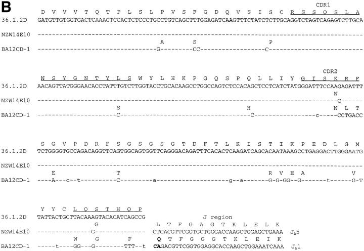
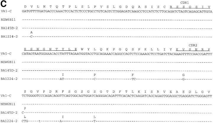
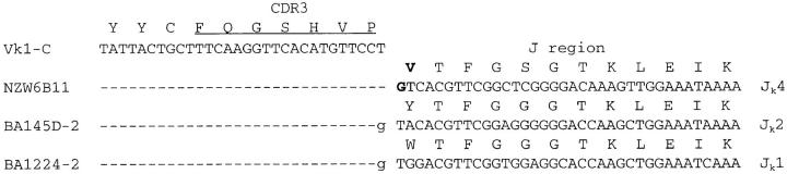
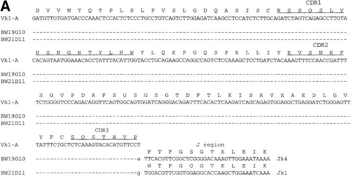
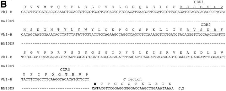
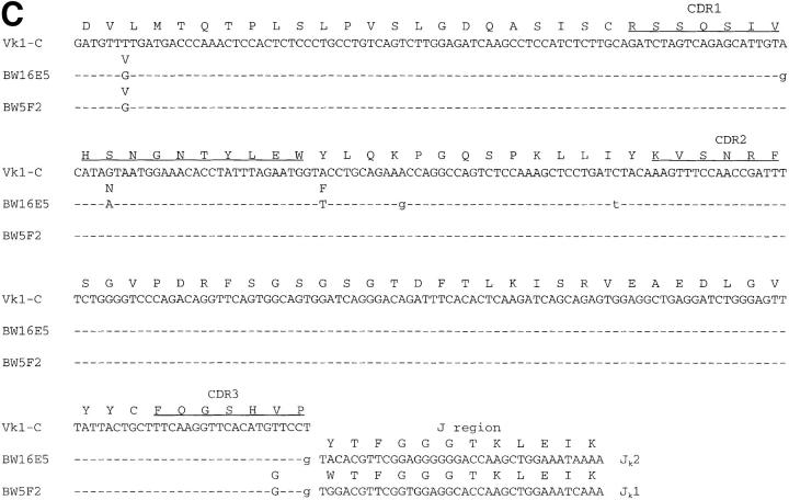
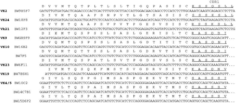
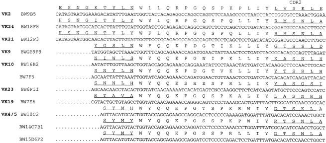
Two major mechanisms for the regulation of autoreactive B cells that arise in the bone marrow are functional silencing (anergy) and deletion. Studies to date suggest that low avidity interactions between B cells and autoantigen lead to B cell silencing, whereas high avidity interactions lead to deletion. Anti-double stranded (ds) DNA antibodies represent a pathogenic autospecificity in Systemic Lupus Erythematosus (SLE). An understanding of their regulation is critical to an understanding of SLE. We now demonstrate in a transgenic model in which mice express the heavy chain of a potentially pathogenic anti-DNA antibody that antibody affinity for dsDNA does not alone determine the fate of anti-dsDNA B cells. B cells making antibodies with similar affinities for dsDNA are regulated differently, depending on light chain usage. A major implication of this observation is that dsDNA may not be the self antigen responsible for cell fate determinations of anti-dsDNA B cells. Light chain usage may determine antigenic cross-reactivity, and cross-reactive antigens may regulate B cells that also bind dsDNA.


Figures

Figure 1
Figure 1
Measurement of R4A-γ2b anti-dsDNA antibody in the sera of NZB/W F1 transgenic and non transgenic mice. Sera from 10 transgenic (Tg+) NZB/W F1 mice and 10 nontransgenic (Tg−) littermates were diluted 1/500 and then tested by ELISA for binding of γ2b antibody to dsDNA. Ages of Tg+ and Tg− mice range from 10–21 wk. There is no linear relationship between age of the mouse and anti-dsDNA titer within these populations. *Mean value significantly exceeds mean value for Tg− mice. P <0.002 (Student's t test).
Figure 2
Figure 2
Nucleotide and amino acid sequences of Vk1 light chains from nonautoimmune transgenic hybridomas. Light chain nucleotide and amino acid sequences from hybridomas from nonautoimmune BALB/c and NZW transgenic mice are compared to their most homologous Vk1 germline genes. (A) Vk1-A; (B) 36.1.2D, an atypical Vk1-α (–6) dextran germline gene; and (C) Vk1-C (–40). CDR regions are underlined and are according to Kabat et al. (40). Dashes indicate identity of nucleotides to the germline sequence. Silent base changes are designated by lowercase letters. Nucleotide changes leading to amino acid replacements in the V region are indicated by uppercase letters. Those leading to amino acid replacements in the joining (J) regions are indicated in bold uppercase letters. Amino acid replacements are shown above the nucleotide sequence. Amino acid replacements from the germline J are indicated in bold. Light chains derived from BALB/c mice have the prefix BA preceding the numeric designation. Light chains derived from NZW mice have the prefix NZW preceding the numeric designation.
Figure 2
Figure 2
Nucleotide and amino acid sequences of Vk1 light chains from nonautoimmune transgenic hybridomas. Light chain nucleotide and amino acid sequences from hybridomas from nonautoimmune BALB/c and NZW transgenic mice are compared to their most homologous Vk1 germline genes. (A) Vk1-A; (B) 36.1.2D, an atypical Vk1-α (–6) dextran germline gene; and (C) Vk1-C (–40). CDR regions are underlined and are according to Kabat et al. (40). Dashes indicate identity of nucleotides to the germline sequence. Silent base changes are designated by lowercase letters. Nucleotide changes leading to amino acid replacements in the V region are indicated by uppercase letters. Those leading to amino acid replacements in the joining (J) regions are indicated in bold uppercase letters. Amino acid replacements are shown above the nucleotide sequence. Amino acid replacements from the germline J are indicated in bold. Light chains derived from BALB/c mice have the prefix BA preceding the numeric designation. Light chains derived from NZW mice have the prefix NZW preceding the numeric designation.
Figure 3
Figure 3
Nucleotide and amino acid sequences of Vk1 light chains from autoimmune transgenic hybridomas. Light chain nucleotide and amino acid sequences from hybridomas from autoimmune NZB/W F1 transgenic mice are compared to their most homologous Vk1 germline genes. (A) Vk1-A, (B) Vk1-B, and (C) Vk1-C (38, 41). CDR regions are underlined and are according to Kabat et al. (40). Dashes indicate identity of nucleotides to the germline sequence. Silent base changes are designated by lowercase letters. Nucleotide changes leading to amino acid replacements in the V region are indicated by uppercase letters. Those leading to amino acid replacements in the J regions are indicated in bold. Amino acid replacements are indicated above the nucleotide sequence. Amino acid replacements from the germline J are indicated in bold. The BW prefix precedes all numeric designations of light chains derived from NZB/W F1 mice.
Figure 3
Figure 3
Nucleotide and amino acid sequences of Vk1 light chains from autoimmune transgenic hybridomas. Light chain nucleotide and amino acid sequences from hybridomas from autoimmune NZB/W F1 transgenic mice are compared to their most homologous Vk1 germline genes. (A) Vk1-A, (B) Vk1-B, and (C) Vk1-C (38, 41). CDR regions are underlined and are according to Kabat et al. (40). Dashes indicate identity of nucleotides to the germline sequence. Silent base changes are designated by lowercase letters. Nucleotide changes leading to amino acid replacements in the V region are indicated by uppercase letters. Those leading to amino acid replacements in the J regions are indicated in bold. Amino acid replacements are indicated above the nucleotide sequence. Amino acid replacements from the germline J are indicated in bold. The BW prefix precedes all numeric designations of light chains derived from NZB/W F1 mice.
Figure 4
Figure 4
Nucleotide and amino acid sequences of non-Vk1 light chains from autoimmune transgenic hybridomas. Light chain nucleotide and amino acid sequences from hybridomas from the autoimmune NZB/W F1 transgenic mice are grouped according to families. Family assignment is based on having >90% homology with other family members as reported in EMBL/GenBank/DDBJ. CDR regions are underlined and are according to Kabat et al. (40). Dots have been introduced in CDR1 to maximize homology. Nucleotide changes leading to amino acid replacements in the J regions are indicated in bold uppercase letters. Amino acids are designated by uppercase letters. Replacements from the germline J region are in bold. N, represents a nucleotide that could not be resolved by sequencing.
Figure 4
Figure 4
Nucleotide and amino acid sequences of non-Vk1 light chains from autoimmune transgenic hybridomas. Light chain nucleotide and amino acid sequences from hybridomas from the autoimmune NZB/W F1 transgenic mice are grouped according to families. Family assignment is based on having >90% homology with other family members as reported in EMBL/GenBank/DDBJ. CDR regions are underlined and are according to Kabat et al. (40). Dots have been introduced in CDR1 to maximize homology. Nucleotide changes leading to amino acid replacements in the J regions are indicated in bold uppercase letters. Amino acids are designated by uppercase letters. Replacements from the germline J region are in bold. N, represents a nucleotide that could not be resolved by sequencing.
Figure 5
Figure 5
Binding to dsDNA. Hybridoma supernatants from BALB/c (BA), NZW, and NZB/W F1 (BW) transgenic mice were normalized to a γ2b concentration of 10 μg/ml and assayed for binding to salmon sperm dsDNA by ELISA. Wildtype R4A-γ2b antibody and irrelevant γ2b are used as positive and negative controls, respectively. Shaded boxes, Vk1 antibodies; open boxes, non-Vk1 antibodies. (A) Strong dsDNAbinding antibodies. Strong dsDNA binders are defined as those whose ELISA measurements at 405 nm are at least fourfold greater than R4A after a 4-h incubation with alkaline phosphatase substrate solution. (B) Moderate dsDNA-binding antibodies. Moderate dsDNA binders are defined as those whose ELISA measurements at 405 nm range from 0.75–2-fold that of R4A after overnight incubation with substrate solution. Results are the average of triplicates ± standard deviation. Standard deviations <0.010 do not appear on the bar graph.
Figure 5
Figure 5
Binding to dsDNA. Hybridoma supernatants from BALB/c (BA), NZW, and NZB/W F1 (BW) transgenic mice were normalized to a γ2b concentration of 10 μg/ml and assayed for binding to salmon sperm dsDNA by ELISA. Wildtype R4A-γ2b antibody and irrelevant γ2b are used as positive and negative controls, respectively. Shaded boxes, Vk1 antibodies; open boxes, non-Vk1 antibodies. (A) Strong dsDNAbinding antibodies. Strong dsDNA binders are defined as those whose ELISA measurements at 405 nm are at least fourfold greater than R4A after a 4-h incubation with alkaline phosphatase substrate solution. (B) Moderate dsDNA-binding antibodies. Moderate dsDNA binders are defined as those whose ELISA measurements at 405 nm range from 0.75–2-fold that of R4A after overnight incubation with substrate solution. Results are the average of triplicates ± standard deviation. Standard deviations <0.010 do not appear on the bar graph.
Figure 6
Figure 6
Glomerular deposition of Vk1 and non-Vk1 antidsDNA antibodies. SCID mice were injected with 5 × 10⁶ hybridoma cells secreting (A) a control non–DNA-binding IgG antibody, (B) aVk1 anti-dsDNA antibody, NZW145D-2, and (C) a non-Vk1 anti-dsDNA antibody, BW 7E6H1. Kidney sections were stained with goat anti–mouse IgG conjugated to alkaline phosphatase and viewed on a Zeiss microscope. ×100. No deposition is observed in mice receiving the cell line producing the non-DNA binding antibody. Glomerular deposits are observed in B and C.

Similar articles

Cited by

References

    1. Gavalchin J, Nicklas JA, Eastcott JW, Madaio MP, Stollar BD, Schwartz RS, Datta SK. Lupus prone (SWR × NZB) F1 mice produce potentially nephritogenic autoantibodies inherited from the normal SWR parent. J Immunol. 1995;134:885–894. - PubMed
    1. Datta SK, Stollar BD, Schwartz RS. Normal mice express idiotypes related to autoantibody idiotypes from lupus mice. Proc Natl Acad Sci USA. 1983;80:2723–2727. - PMC - PubMed
    1. Krishnan MR, Marion TN. Structural similarity of antibody variable regions from immune and autoimmune anti-DNA antibodies. J Immunol. 1993;150:4948–4957. - PubMed
    1. Kofler R, Noonan DJ, Levy DE, Wilson MC, Moller NA, Dixon FJ, Theofilopoulos AN. Genetic elements used for a murine lupus anti-DNA autoantibody are closely related to those for antibodies to exogenous antigens. J Exp Med. 1985;161:805–815. - PMC - PubMed
    1. Manheimer-Lory A, Monhian R, Splaver A, Gaynor B, Diamond B. Analysis of the Vk1 family: germline genes from an SLE patient and expressed autoantibodies. Autoimmunity. 1995;20:259–265. - PubMed

Publication types

MeSH terms