. 1997 Apr 7;185(7):1387-92.
doi: 10.1084/jem.185.7.1387.

Role of tyrosine phosphorylation of HS1 in B cell antigen receptor-mediated apoptosis

Affiliations

Role of tyrosine phosphorylation of HS1 in B cell antigen receptor-mediated apoptosis

Y Yamanashi et al. J Exp Med. .

Abstract

The 75-kD HS1 protein is highly tyrosine-phosphorylated during B cell antigen receptor (BCR)-mediated signaling. Owing to low expression of HS1, WEHI-231-derived M1 cells, unlike the parental cells, are insensitive to BCR-mediated apoptosis. Here, we show that BCR-associated tyrosine kinases Lyn and Syk synergistically phosphorylate HS1, and that Tyr-378 and Tyr-397 of HS1 are the critical residues for its BCR-induced phosphorylation. In addition, unlike wild-type HS1, a mutant HS1 carrying the mutations Phe-378 and Phe-397 was unable to render M1 cells sensitive to apoptosis. Wild-type HS1, but not the mutant, localized to the nucleus under the synergy of Lyn and Syk. Thus, tyrosine phosphorylation of HS1 is required for BCR-induced apoptosis and nuclear translocation of HS1 may be a prerequisite for B cell apoptosis.


Figures

Figure 1
Figure 1
Cooperation between Lyn and Syk in HS1 phosphorylation. CV-1 cells (1.5 × 10⁶) were transfected with the expression plasmids (5 μg), pMELyn (+), pME-Syk (+), pMEHS1 (+), or with H2O in place of DNA solution (−), and lysed with TNE buffer. The lysates were subjected to immunoprecipitation with anti-serum to HS1. The immunoprecipitates were immunoblotted with PY20 (top). Aliquots (1/50) of the lysates were immunoblotted with anti-HS1 Ab (bottom). Positions of HS1 and standard protein markers (kD) are indicated.
Figure 2
Figure 2
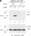
Requirement of Tyr-378 and Tyr-397 in tyrosine phosphorylation of HS1 upon BCR stimulation. (A) Schematic structure of the human HS1 protein. The 23 amino acids sequence of HS1, Tyr-378 to Val-400, deleted to generate HS1-ΔYY, is shown on the top. Possible target sites of the SH2 domain of the Src-like kinase are underlined. Two tyrosine residues, Tyr-378 and Tyr-397, in the sequence are indicated in bold. The positions of the two tyrosine residues (YY), three helix–turn– helix repeats (HTH repeats) and the SH3 domain (SH3) are indicated in the schematic drawing of HS1. The amino acid numbers (from Met-1 to Glu-486) are shown on the bottom. (B) Tyrosine phosphorylation of HS1 and HS1-ΔYY in CV-1 cells. CV-1 cells (1.5 × 10⁶) were transfected with the expression plasmids (5 μg), pME-Lyn, pME-Syk, and pMEHS1-ΔYY (ΔYY) or pME-HS1 (HS1), then lysed with TNE buffer. The lysates were equally divided into two samples and subjected to immunoprecipitation with antiserum to HS1. The immunoprecipitates were analyzed by anti-HS1 (HS1) or by PY-20 immunoblotting. The levels of Lyn and/or Syk-mediated tyrosine phosphorylation of the other cellular proteins were virtually the same between the cells expressing wild-type HS1 and those expressing HS1-ΔYY (data not shown). Positions of HS1, HS1-ΔYY, and a 96-kD standard protein marker are indicated. (C) Tyrosine phosphorylation of HS1 mutants in WEHI-231 cells. WEHI-231derived cells (1 × 10⁷), which express human HS1 (lanes 1 and 2), HS1FF (lanes 3 and 4), HS1-FY (lanes 5 and 6), or HS1-YF (lanes 7 and 8), were incubated with (lanes 2, 4, 6, and 8) or without (lanes 1, 3, 5, and 7) anti-IgM Ab. The cell lysates were subjected to immunoprecipitation with the human HS1-specific Ab and the immunoprecipitates probed by anti-phosphotyrosine (PY blot) or anti-human HS1 (hHS1 blot) immunoblotting. BCRmediated tyrosine phosphorylation of the other proteins was not affected by expressing exogenous HS1 or its mutants (data not shown). Positions of HS1 and standard protein markers are indicated.
Figure 2
Figure 2
Requirement of Tyr-378 and Tyr-397 in tyrosine phosphorylation of HS1 upon BCR stimulation. (A) Schematic structure of the human HS1 protein. The 23 amino acids sequence of HS1, Tyr-378 to Val-400, deleted to generate HS1-ΔYY, is shown on the top. Possible target sites of the SH2 domain of the Src-like kinase are underlined. Two tyrosine residues, Tyr-378 and Tyr-397, in the sequence are indicated in bold. The positions of the two tyrosine residues (YY), three helix–turn– helix repeats (HTH repeats) and the SH3 domain (SH3) are indicated in the schematic drawing of HS1. The amino acid numbers (from Met-1 to Glu-486) are shown on the bottom. (B) Tyrosine phosphorylation of HS1 and HS1-ΔYY in CV-1 cells. CV-1 cells (1.5 × 10⁶) were transfected with the expression plasmids (5 μg), pME-Lyn, pME-Syk, and pMEHS1-ΔYY (ΔYY) or pME-HS1 (HS1), then lysed with TNE buffer. The lysates were equally divided into two samples and subjected to immunoprecipitation with antiserum to HS1. The immunoprecipitates were analyzed by anti-HS1 (HS1) or by PY-20 immunoblotting. The levels of Lyn and/or Syk-mediated tyrosine phosphorylation of the other cellular proteins were virtually the same between the cells expressing wild-type HS1 and those expressing HS1-ΔYY (data not shown). Positions of HS1, HS1-ΔYY, and a 96-kD standard protein marker are indicated. (C) Tyrosine phosphorylation of HS1 mutants in WEHI-231 cells. WEHI-231derived cells (1 × 10⁷), which express human HS1 (lanes 1 and 2), HS1FF (lanes 3 and 4), HS1-FY (lanes 5 and 6), or HS1-YF (lanes 7 and 8), were incubated with (lanes 2, 4, 6, and 8) or without (lanes 1, 3, 5, and 7) anti-IgM Ab. The cell lysates were subjected to immunoprecipitation with the human HS1-specific Ab and the immunoprecipitates probed by anti-phosphotyrosine (PY blot) or anti-human HS1 (hHS1 blot) immunoblotting. BCRmediated tyrosine phosphorylation of the other proteins was not affected by expressing exogenous HS1 or its mutants (data not shown). Positions of HS1 and standard protein markers are indicated.
Figure 3
Figure 3
HS1 tyrosine phosphorylation is required for BCR-mediated apoptosis. (A) Restoration of BCR-mediated apoptosis by exogenous HS1 but not by HS1-FF in M1 cells. WEHI-231 cells (1 and 2), M1 cells (3 and 4), M1-derived cells expressing wild-type HS1 (5 and 6), and M1-derived cells expressing the HS1-FF protein (7 and 8) (1–2 × 10⁵) were incubated for 48 h with (2, 4, 6, and 8) or without (1, 3, 5, and 7) anti-IgM Ab, stained with propidium iodide, and analyzed by flow cytometry. The DNA content of the cells (non-gated) was expressed as the histogram of propidium iodide fluorescence intensity. The percentage of apoptotic cells is denoted in each histogram (above horizontal bars). The results are representative of three independent experiments. Cells containing subdiploid DNA represent apoptotic cells. (B) Expression and tyrosine phosphorylation of wild-type HS1 and HS1-FF in M1 cells. M1-derived cells (1 × 10⁷), which express the human HS1 (lanes 1 and 2) or HS1-FF (lanes 3 and 4), were incubated with (lanes 2 and 4) or without (lanes 1 and 3) anti-IgM Ab. The cells were then lysed in TNE buffer and the lysate were analyzed by immunoprecipitation with the anti-HS1 mAb and by anti-phosphotyrosine (PY blot) or antihuman HS1 (hHS1 blot) immunoblotting of the immunoprecipitates. Positions of HS1 and standard protein markers are indicated.
Figure 3
Figure 3
HS1 tyrosine phosphorylation is required for BCR-mediated apoptosis. (A) Restoration of BCR-mediated apoptosis by exogenous HS1 but not by HS1-FF in M1 cells. WEHI-231 cells (1 and 2), M1 cells (3 and 4), M1-derived cells expressing wild-type HS1 (5 and 6), and M1-derived cells expressing the HS1-FF protein (7 and 8) (1–2 × 10⁵) were incubated for 48 h with (2, 4, 6, and 8) or without (1, 3, 5, and 7) anti-IgM Ab, stained with propidium iodide, and analyzed by flow cytometry. The DNA content of the cells (non-gated) was expressed as the histogram of propidium iodide fluorescence intensity. The percentage of apoptotic cells is denoted in each histogram (above horizontal bars). The results are representative of three independent experiments. Cells containing subdiploid DNA represent apoptotic cells. (B) Expression and tyrosine phosphorylation of wild-type HS1 and HS1-FF in M1 cells. M1-derived cells (1 × 10⁷), which express the human HS1 (lanes 1 and 2) or HS1-FF (lanes 3 and 4), were incubated with (lanes 2 and 4) or without (lanes 1 and 3) anti-IgM Ab. The cells were then lysed in TNE buffer and the lysate were analyzed by immunoprecipitation with the anti-HS1 mAb and by anti-phosphotyrosine (PY blot) or antihuman HS1 (hHS1 blot) immunoblotting of the immunoprecipitates. Positions of HS1 and standard protein markers are indicated.
Figure 4
Figure 4
Subcellular localization of HS1. (A) Nuclear localization of HS1 coexpressed with Lyn and Syk. COS7 cells (1.5 × 10⁶) were transfected with the expression plasmids (5 μg), pME-Lyn, pME-Syk, and either pME-HS1 (b), or pME-HS1-FF (c). As a control, cells were transfected with pME-HS1 alone (a). Subcellular localization of the HS1 protein was investigated by indirect immunofluorescence microscopy using antiHS1 mAb and FITC-labeled second Ab. No significant signal was detectable in the absence of primary Ab and in untransfected COS7 cells. (B) Increment of nuclear HS1 in BCR-stimulated WEHI-231 cells. WEHI-231 cells were incubated with anti-IgM Ab for the time indicated above. Then the cells were subjected to subcellular fractionation. Samples of nuclear fraction from 2 × 10⁵ cells per lane were tested for HS1 by immunoblotting.
Figure 4
Figure 4
Subcellular localization of HS1. (A) Nuclear localization of HS1 coexpressed with Lyn and Syk. COS7 cells (1.5 × 10⁶) were transfected with the expression plasmids (5 μg), pME-Lyn, pME-Syk, and either pME-HS1 (b), or pME-HS1-FF (c). As a control, cells were transfected with pME-HS1 alone (a). Subcellular localization of the HS1 protein was investigated by indirect immunofluorescence microscopy using antiHS1 mAb and FITC-labeled second Ab. No significant signal was detectable in the absence of primary Ab and in untransfected COS7 cells. (B) Increment of nuclear HS1 in BCR-stimulated WEHI-231 cells. WEHI-231 cells were incubated with anti-IgM Ab for the time indicated above. Then the cells were subjected to subcellular fractionation. Samples of nuclear fraction from 2 × 10⁵ cells per lane were tested for HS1 by immunoblotting.

Similar articles

Cited by

References

    1. Yamanashi Y, Kakiuchi T, Mizuguchi J, Yamamoto T, Toyoshima K. Association of B cell antigen receptor with protein tyrosine kinase Lyn. Science (Wash DC) 1991;251:192–194. - PubMed
    1. Bolen JB. Protein tyrosine kinases in the initiation of antigen receptor signaling. Curr Opin Immunol. 1995;7:306–311. - PubMed
    1. Hibbs ML, Tarlinton DM, Armes J, Grail D, Hodgson G, Maglitto R, Stacker SA, Dunn AR. Multiple defects in the immune system of Lyn-deficient mice, culminating in autoimmune disease. Cell. 1995;83:301–311. - PubMed
    1. Nishizumi H, Taniuchi I, Yamanashi Y, Kitamura D, Ilic D, Mori S, Watanabe T, Yamamoto T. Impaired proliferation of peripheral B cells and indication of autoimmune disease in lyn-deficient mice. Immunity. 1995;3:549–560. - PubMed
    1. Takata M, Sabe H, Hata A, Inazu T, Homma Y, Nukada T, Yamamura H, Kurosaki T. Tyrosine kinase Lyn and Syk regulates B cell receptor-coupled Ca2+mobilization through distinct pathways. EMBO (Eur Mol Biol Organ) J. 1994;13:1341–1349. - PMC - PubMed

Publication types