. 1997 Apr 7;137(1):19-26.
doi: 10.1083/jcb.137.1.19.

High mobility group 1 protein is not stably associated with the chromosomes of somatic cells

Affiliations

High mobility group 1 protein is not stably associated with the chromosomes of somatic cells

L Falciola et al. J Cell Biol. .

Abstract

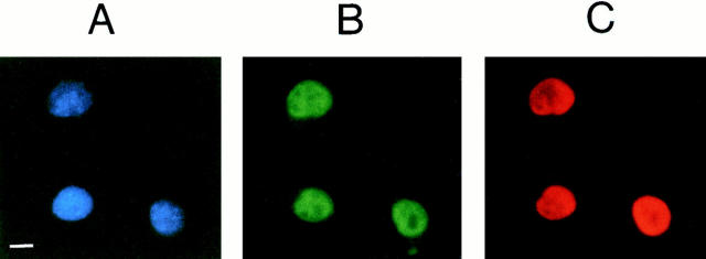
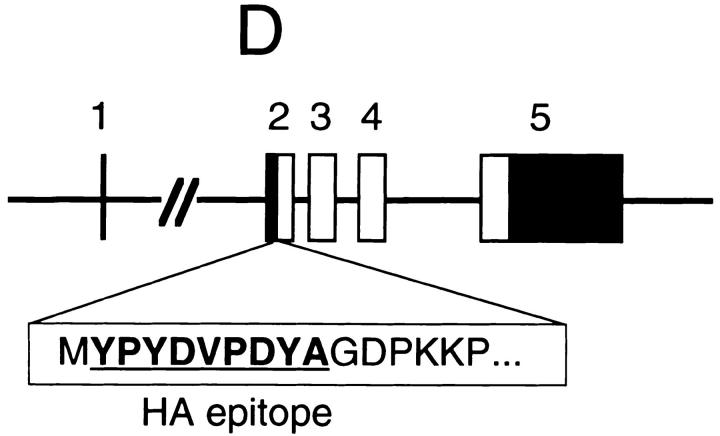
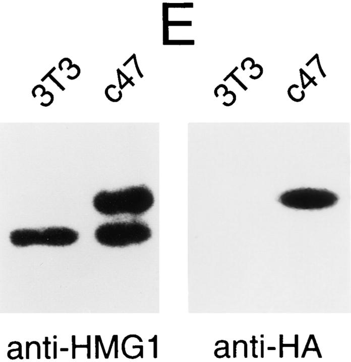
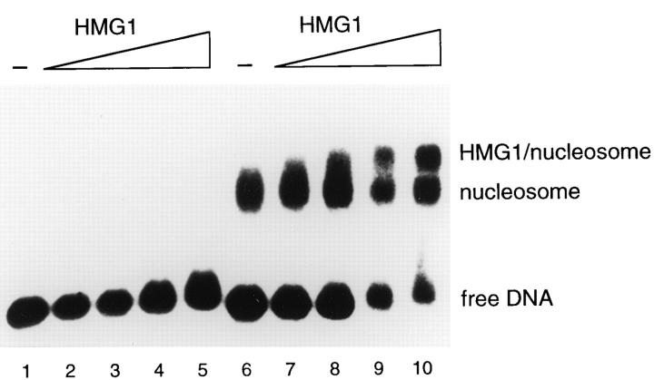
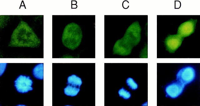
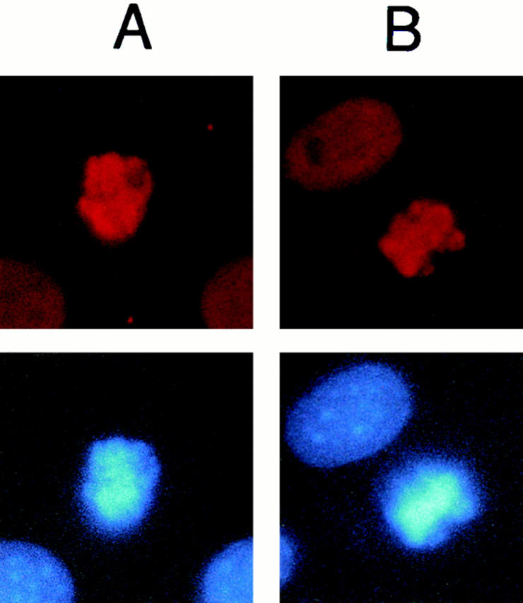
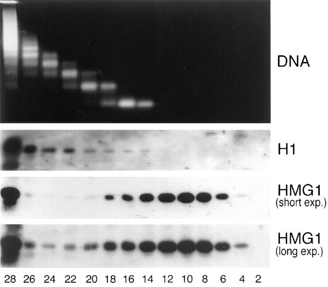
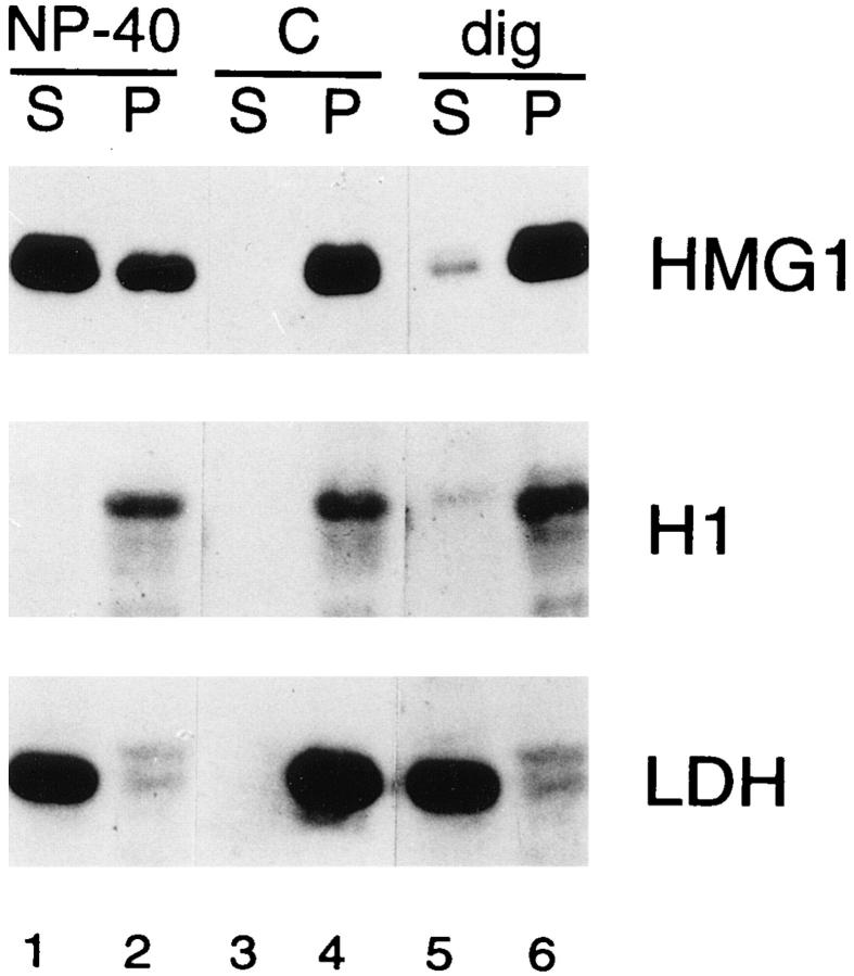
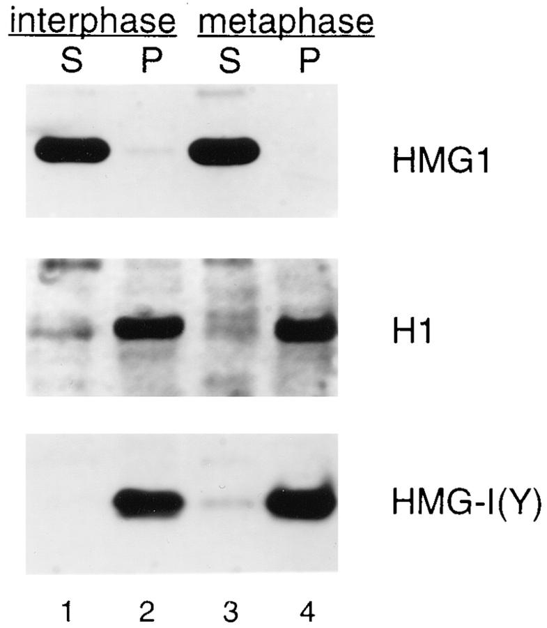
High mobility group 1 (HMG1) protein is an abundant and conserved component of vertebrate nuclei and has been proposed to play a structural role in chromatin organization, possibly similar to that of histone H1. However, a high abundance of HMG1 had also been reported in the cytoplasm and on the surface of mammalian cells. We conclusively show that HMG1 is a nuclear protein, since several different anti-HMG1 antibodies stain the nucleoplasm of cultured cells, and epitope-tagged HMG1 is localized in the nucleus only. The protein is excluded from nucleoli and is not associated to specific nuclear structures but rather appears to be uniformly distributed. HMG1 can bind in vitro to reconstituted core nucleosomes but is not stably associated to chromatin in live cells. At metaphase, HMG1 is detached from condensed chromosomes, contrary to histone H1. During interphase, HMG1 readily diffuses out of nuclei after permeabilization of the nuclear membranes with detergents, whereas histone H1 remains associated to chromatin. These properties exclude a shared function for HMG1 and H1 in differentiated cells, in spite of their similar biochemical properties. HMG1 may be stably associated only to a very minor population of nucleosomes or may interact transiently with nucleosomes during dynamic processes of chromatin remodeling.


Figures

Figure 1
Figure 1
(A) Reactivity of the various anti-HMG1 antibodies used in this study. For each antibody preparation the species of origin is indicated (m, mouse; ch, chicken), and the ability (+) or inability (−) to recognize boxes A and B of HMG1 by Western blotting (denatured) or by immunoprecipitation (native) is also shown. The notation ± indicates that recovery of the HMG1bA polypeptide by immunoprecipitation is <5% under the conditions indicated under Materials and Methods. (B) Reactivity of the chWB-AB antibody in Western blots. Whole NIH 3T3 cells were lysed by addition of SDSPAGE loading buffer, and 10 μg of total protein was loaded on a 10% tricine–SDS– polyacrylamide gel (lane 3T3), alongside 20 ng of purified recombinant HMG1bA polypeptide (lane bA) or 20 ng of purified recombinant HMG1bB polypeptide (lane bB). (C and D) Anti-HMG1 antibodies stain the cell nucleus. NIH 3T3 cells were grown on glass coverslips, fixed with paraformaldehyde, permeabilized with 0.1% NP-40, and stained with anti-recAtn (C) and Hoechst 33258 (D). (E and F) Localization of HMG1 by confocal microscopy. HeLa cells were fixed with paraformaldehyde, permeabilized with 0.1% SDS, stained with mAP-bA antibody, and viewed in green fluorescence (E) or by phase contrast microscopy (F). Bars, 10 μm.
Figure 1
Figure 1
(A) Reactivity of the various anti-HMG1 antibodies used in this study. For each antibody preparation the species of origin is indicated (m, mouse; ch, chicken), and the ability (+) or inability (−) to recognize boxes A and B of HMG1 by Western blotting (denatured) or by immunoprecipitation (native) is also shown. The notation ± indicates that recovery of the HMG1bA polypeptide by immunoprecipitation is <5% under the conditions indicated under Materials and Methods. (B) Reactivity of the chWB-AB antibody in Western blots. Whole NIH 3T3 cells were lysed by addition of SDSPAGE loading buffer, and 10 μg of total protein was loaded on a 10% tricine–SDS– polyacrylamide gel (lane 3T3), alongside 20 ng of purified recombinant HMG1bA polypeptide (lane bA) or 20 ng of purified recombinant HMG1bB polypeptide (lane bB). (C and D) Anti-HMG1 antibodies stain the cell nucleus. NIH 3T3 cells were grown on glass coverslips, fixed with paraformaldehyde, permeabilized with 0.1% NP-40, and stained with anti-recAtn (C) and Hoechst 33258 (D). (E and F) Localization of HMG1 by confocal microscopy. HeLa cells were fixed with paraformaldehyde, permeabilized with 0.1% SDS, stained with mAP-bA antibody, and viewed in green fluorescence (E) or by phase contrast microscopy (F). Bars, 10 μm.
Figure 2
Figure 2
The product of the Hmg1 gene localizes to the nucleus. NIH 3T3 fibroblasts stably transfected with the pHMG1tag plasmid were fixed and stained simultaneously with (A) Hoechst 33258, (B) anti-HMG1 antibody chIPAB, and (C) monoclonal antibody 12CA5 recognizing the HA epitope. (D) Structure of the Hmg1-tag gene. The mouse gene Hmg1, which codes for protein HMG1, is transcribed under the control of its own strong promoter/enhancer. Exons are numbered. Black boxes represent untranslated sequences and white boxes translated sequences. Plasmid pmHMG1 was modified by the insertion of 27 bp coding for the HA epitope (bold and underlined) immediately after the ATG codon for the first methionine of HMG1 and in frame with the rest of the protein. A stable clone (c47) expressing HMG1tag approximately to the same level of unmodified HMG1 was selected. (E) Western blotting of whole cell extracts from wild-type fibroblasts (lanes 3T3) and from the stable transfected cells (lanes c47). The anti-HMG1 antibody chWB-AB (left) recognizes in the same way HMG1 and HMG1tag; the monoclonal antibody 12CA5 recognizes the HA epitope (right). Protein HMG1tag runs slightly slower than wild-type HMG1 in tricine– SDS-PAGE because of the addition of nine amino acids.
Figure 2
Figure 2
The product of the Hmg1 gene localizes to the nucleus. NIH 3T3 fibroblasts stably transfected with the pHMG1tag plasmid were fixed and stained simultaneously with (A) Hoechst 33258, (B) anti-HMG1 antibody chIPAB, and (C) monoclonal antibody 12CA5 recognizing the HA epitope. (D) Structure of the Hmg1-tag gene. The mouse gene Hmg1, which codes for protein HMG1, is transcribed under the control of its own strong promoter/enhancer. Exons are numbered. Black boxes represent untranslated sequences and white boxes translated sequences. Plasmid pmHMG1 was modified by the insertion of 27 bp coding for the HA epitope (bold and underlined) immediately after the ATG codon for the first methionine of HMG1 and in frame with the rest of the protein. A stable clone (c47) expressing HMG1tag approximately to the same level of unmodified HMG1 was selected. (E) Western blotting of whole cell extracts from wild-type fibroblasts (lanes 3T3) and from the stable transfected cells (lanes c47). The anti-HMG1 antibody chWB-AB (left) recognizes in the same way HMG1 and HMG1tag; the monoclonal antibody 12CA5 recognizes the HA epitope (right). Protein HMG1tag runs slightly slower than wild-type HMG1 in tricine– SDS-PAGE because of the addition of nine amino acids.
Figure 3
Figure 3
HMG1 binds to reconstituted mononucleosomes. A labeled DNA fragment (176 bp) was assembled into mononucleosomes, incubated with increasing amounts (0, 10, 50, 100, and 500 ng) of HMG1 (lanes 6–10), and electrophoresed on a 0.7% agarose gel. DNA not assembled in nucleosomes was treated similarly for comparison (lanes 1–5). The bands corresponding to free DNA, to nucleosome particles, and to HMG1–nucleosome complexes are indicated.
Figure 4
Figure 4
HMG1 protein is not associated to mitotic condensed chromosomes. Dividing NIH 3T3 fibroblasts were fixed and stained for HMG1 with antibody chIP-AB (top row, green fluorescence) and for DNA with Hoechst 33258 (bottom row, blue fluorescence). Representative cells at different stages during mitosis: prophase (A), metaphase (B), anaphase (C), and telophase (D). After the breakdown of the nuclear membrane, HMG1 diffuses throughout the cytoplasm, and the pattern of green fluorescence corresponds to the shape of the cell. However, fluorescence from HMG1 is clearly reduced in correspondence to the volume occupied by condensed chromosomes (compare top and bottom images), indicating that HMG1 is not associated with DNA during mitosis. After cell division and the reformation of nuclear membranes (D), the majority of HMG1 colocalizes with DNA, but some is still found in the cytoplasm, suggesting that the protein is being concentrated in the nuclei by passage through the nuclear membrane.
Figure 5
Figure 5
Core histone H2B and linker histone H1 remain associated to condensed chromosomes throughout mitosis. As a control for the mitotic displacement observed with HMG1, dividing NIH 3T3 fibroblasts were fixed and stained for DNA with Hoechst 33258 (bottom) and either (A) for histone H2B with the monoclonal IgM HBC-7 (top) or (B) for linker histones with a rabbit polyclonal antibody against histone H1 (top).
Figure 6
Figure 6
A minor fraction of HMG1 cofractionates with polynucleosomes in sucrose gradients. Nuclei of NIH 3T3 fibroblasts were partially digested with micrococcal nuclease, lysed, and sedimented through a sucrose gradient (see Materials and Methods). Individual fractions were analyzed by Western blotting for the presence of HMG1 and histone H1; DNA was also extracted from the fractions and analyzed on a 2% agarose gel.
Figure 7
Figure 7
HMG1 leaks out from detergent-permeabilized nuclei of adherent cells, but histone H1 does not. Adherent NIH 3T3 fibroblasts were incubated in buffer with no detergent (lanes 3 and 4), in buffer containing 0.1% NP-40 (lanes 1 and 2), or in buffer containing 40 μg/ml digitonin (lanes 5 and 6). After incubation, the buffer bathing the cells (lanes 1, 3, and 5; S, supernatant) and the remnants of permeabilized cells (lanes 2, 4, and 6; P, pellet) were analyzed by Western blotting with antibodies against LDH, HMG1, and histone H1. Digitonin selectively permeabilizes the plasma membrane and causes the complete leakage of LDH but not of HMG1 and H1. The faint bands in lanes 2 and 6 do not correspond to LDH, because they have a slightly different molecular weight. NP-40 causes the disruption of all membranes including the nuclear ones: LDH is completely released, as well as ∼75% of HMG1 (as determined by densitometric analysis), but H1 remains associated with the DNA. We observed an incomplete release of HMG1 only when we permeabilized cells still attached to their plastic substrate, possibly because HMG1 sticks avidly to secreted glycoproteins (Falciola, L., and M.E. Bianchi, unpublished results).
Figure 8
Figure 8
HMG1 leaks out in a similar way from both interphasic and mitotic permeabilized cells. NIH 3T3 fibroblasts were exposed overnight to nocodazole, an inhibitor of microtubule polymerization. Cells that had entered M phase could not proceed further and were detached from their plastic substrate by manual shaking (metaphase cells). Cells that remained adherent to the substrate after shaking (interphase cells) were detached by treatment with trypsin. The two cell populations were checked for the presence of condensed chromosomes (95% for metaphase cells; 2% for interphase cells). The cell suspensions were then exposed to 0.1% NP-40 and immediately centrifuged. Supernatants (lanes 1 and 3, S) and cell pellets (lanes 2 and 4, P) were analyzed by Western blotting with antibodies against HMG1, histone H1, and protein HMG-I(Y).

References

    1. Adam SA, Sterne R, Marr, Gerace L. Nuclear protein import in permeabilized mammalian cells requires soluble cytoplasmic factors. J Cell Biol. 1990;111:807–816. - PMC - PubMed
    1. Bianchi ME. Prokaryotic HU and eukaryotic HMG1: a kinked relationship. Mol Microbiol. 1994;14:1–5. - PubMed
    1. Bianchi, M.E. 1995. The HMG-box domain. In DNA–Protein: Structural Interactions. D.M.J. Lilley, editor. Oxford University Press, Oxford. 177–200.
    1. Bianchi ME, Beltrame M, Paonessa G. Specific recognition of cruciform DNA by nuclear protein HMG1. Science (Wash DC) 1989;243:1056–1059. - PubMed
    1. Bianchi, M.E., L. Falciola, S. Ferrari, and D.M.J. Lilley. 1992. The DNA binding site of HMG1 protein is composed of two similar segments (HMG boxes), both of which have counterparts in other eukaryotic regulatory proteins. EMBO (Eur. Mol. Biol. Organ.) J. 11:1055–1063. - PMC - PubMed

Publication types

MeSH terms