. 1997 Apr 7;137(1):37-49.
doi: 10.1083/jcb.137.1.37.

Chromatin degradation in differentiating fiber cells of the eye lens

Affiliations

Chromatin degradation in differentiating fiber cells of the eye lens

S Bassnett et al. J Cell Biol. .

Abstract

During development, the lens of the eye becomes transparent, in part because of the elimination of nuclei and other organelles from the central lens fiber cells by an apoptotic-like mechanism. Using confocal microscopy we showed that, at the border of the organelle-free zone (OFZ), fiber cell nuclei became suddenly irregular in shape, with marginalized chromatin. Subsequently, holes appeared in the nuclear envelope and underlying laminae, and the nuclei collapsed into condensed, spherical structures. Nuclear remnants, containing DNA, histones, lamin B2, and fragments of nuclear membrane, were detected deep in the OFZ. We used in situ electrophoresis to demonstrate that fragmented DNA was present only in cells bordering the OFZ. Confocal microscopy of terminal deoxynucleotidyl transferase (TdT)-labeled lens slices confirmed that DNA fragmentation was a relatively late event in fiber differentiation, occurring after the loss of the nuclear membrane. DNA fragments with 3'-OH or 3'-PO(4) ends were not observed elsewhere in the lens under normal conditions, although they could be produced by pretreatment with DNase I or micrococcal nuclease, respectively. Dual labeling with TdT and an antibody against protein disulfide isomerase, an ER-resident protein, revealed a distinct spatial and temporal gap between the disappearance of ER and nuclear membranes and the onset of DNA degradation. Thus, fiber cell chromatin disassembly differs significantly from classical apoptosis, in both the sequence of events and the time course of the process. The fact that DNA degradation occurs only after the disappearance of mitochondrial, ER, and nuclear membranes suggests that damage to intracellular membranes may be an initiating event in nuclear breakdown.


Figures

Figure 1
Figure 1
Diagram of a midsagittal slice of a chicken lens at E15. The lens is bounded by an acellular collagenous capsule. An epithelial monolayer covers the anterior surface of the lens and thickens at the periphery to form the annular pad. The bulk of the lens consists of concentric layers of highly elongated lens fiber cells derived from the edges of the epithelium. Primary fiber cells, formed early in development, are situated in the center of the lens. The rest of the fiber mass is composed of secondary fiber cells, formed throughout life by the differentiation of epithelial cells at the lens equator. The tips of secondary fiber cells make contact with fibers from the opposite hemisphere of the lens at the sutures. The outer fiber cells contain a normal complement of organelles, including nuclei. However, organelles are absent from cells in the center of the lens, giving rise to the OFZ. The OFZ increases steadily in size throughout embryonic development (small arrows).
Figure 2
Figure 2
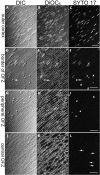
The fate of the nuclear membrane and DNA in fiber cells near the border of the OFZ. The lens slice was stained with DiOC6 (for membranes) and SYTO 17 (for DNA) and viewed with a confocal microscope. Cortical fiber cells are shown in A–C, cells just outside the border of the OFZ are shown in D–F, cells immediately within the OFZ are shown in G–I, and cells in the center of the lens are shown in J–L. A, D, G, and J are differential interference contrast images; B, E, H, and K, are confocal images of the DiOC6 fluorescence; and C, F, I, and L are the corresponding confocal images of the SYTO 17 fluorescence. Note the sudden change in nuclear morphology at the border of the OFZ and the loss of the nuclear membrane (D–F). The last nucleus to possess a nuclear membrane is indicated by the arrow in D–F. Collapsed nuclei are evident in cells within the OFZ and SYTO 17–stained nuclear remnants (L, arrows) can be discerned, even in the most central fiber cells. Bar, 25 μm.
Figure 3
Figure 3
Confocal micrographs of lamin B2 immunofluorescence in an E17 lens slice after high-temperature antigen retrieval (see text for details). (A) Fiber cells in the peripheral cortex. The immunofluorescence is restricted to the lamina of the fiber cell nuclei. (B) Lowmagnification view of cells near the border of the OFZ. Note that even nuclear remnants (arrowhead) are positive for lamin B2 immunofluorescence. (C) High-magnification view of cells immediately adjacent to the OFZ (B, arrow). In many of these cells, the nuclear lamina has become distorted, and the profiles of the nuclei are irregular (arrows), although some nuclei still retain a more normal appearance (arrowhead). (D) High-magnification view of cells immediately within the OFZ (B, arrowhead). At this point in the denucleation process, the fiber nuclei have collapsed into small spherical structures (arrows), and rents are apparent in the nuclear lamina. Bars: (A) 25 μm; (B) 50 μm; (C) 10 μm; (D) 5 μm.
Figure 4
Figure 4
DIC and corresponding confocal immunofluorescence images of histone distribution in an E17 lens slice. The antibody recognizes an epitope common to histones H1, H2A, H2B, H3, and H4. (A) DIC image of the equatorial region of a lens slice showing the annular pad (ap) and superficial fibers. (B) Immunofluorescence image of the region shown in A. Note the strong labeling of nuclei in the annular pad and superficial fibers. (C) DIC image of fiber cells at the border of the OFZ. Fiber cell nuclei at various stages of disassembly are present, including those with marginalized (arrow) or condensed chromatin (arrowhead). (D) Immunofluorescence image of the region shown in C. During fiber cell denucleation, the distribution of histone proteins parallels that of the DNA (compare with Fig. 2 F  ), becoming first marginalized (arrow) and then collapsing into condensed residual structures (arrowhead). Bars, (A and B) 50 μm; (C and D) 10 μm.
Figure 5
Figure 5
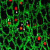
Merged confocal image of fiber cells immediately inside the border of the OFZ after staining with DiOC6 (green) and SYTO 17 (red). The combination of these fluorescent probes allows the simultaneous visualization of the fiber cell membranes (DiOC6) and nuclear DNA (SYTO 17). At this point in fiber cell differentiation, the nuclei have collapsed into condensed spherical structures. Remnants of the nuclear membrane are still visible (arrowheads), attached to the naked chromatin. The highly interdigitated lateral membranes of the fiber cells are also evident. Bar, 5 μm.
Figure 6
Figure 6
Merged confocal and DIC images of lens slices after TdT labeling with fluorescein-dUTP. The DIC images are shown in green and positively labeled nuclei (containing fragmented DNA) are shown in red. (A) At the border of the OFZ, the nuclei lose their regular shape (arrows) and collapse into condensed structures that are strongly labeled by the TdT assay (arrowheads). Positively labeled debris, resulting presumably from the disintegration of labeled nuclei, extends deep into the OFZ. (B) Cortical fiber cells from a lens slice that was pretreated for 30 min with 50 U/ml DNase I. Note that after DNase I treatment, all nuclei are labeled by the TdT assay and that the labeling is strongest immediately beneath the nuclear membrane. (C) Equatorial region of a lens slice that had been incubated with CIAP before TdT labeling. None of the nuclei are labeled, indicating that the superficial fibers do not contain fragmented DNA with 3′PO4 termini (see text for details). (D) Equatorial region of a lens slice that was treated sequentially with micrococcal nuclease and CIAP before TdT labeling. All the nuclei are labeled, demonstrating the efficacy of the CIAP technique for detecting fragmented DNA with 3′-PO4 termini. Bars: (A) 50 μm; (B) 10 μm; (C) 50 μm; (D) 50 μm.
Figure 7
Figure 7
Confocal images of lens slices showing the distribution of ER (green) and degraded DNA (red). (A) The ER (visualized by immunofluorescence with a protein disulfide isomerase antibody) is abundant in the superficial layers of the lens but completely absent from the well-defined central OFZ. The fiber cell nuclear membranes are also labeled by this antibody. Degraded DNA is localized in condensed nuclear remnants, scattered throughout the cytoplasm of fiber cells in the OFZ. Note the gap of ∼90 μm between the last fiber cell to contain ER (arrow) and the first to contain degraded DNA (arrowhead). e, epithelium; ap, annular pad. (B) Apoptotic cells are occasionally detected in the anterior epithelium of the E9 lens. At this stage of development, the epithelial cells (e) and all of the fiber cells ( f ) contain abundant ER. Apoptotic nuclei (arrows) are strongly labeled by the TdT assay, and small apoptotic bodies are often observed in the adjacent tissue (arrowheads). Note that, in contrast to the central fiber cells shown in A, the apoptotic nuclei of the epithelial cells are found in cells in which the ER is still present. Bars: (A) 100 μm; (B) 25 μm.
Figure 8
Figure 8
Diagram showing the principle of in situ electrophoresis. (A) A permeabilized, alkali-treated lens slice is embedded in a block of 0.3% agarose and placed in a weak electric field. Electrophoretically mobile, fragmented DNA is driven from the slice into the gel. The preparation is stained with ethidium bromide and transferred to the stage of a laser scanning confocal microscope (LSM). The focal plane of the microscope is positioned midway up the slice at the level of the fiber cell nuclei. A hypothetical view down the microscope is shown in B, the OFZ appearing as a dark region in a strip of brightly stained nuclei. Fragmented DNA is visualized as streams of positively stained material emanating from regions of the lens where the DNA was sufficiently degraded to be rendered electrophoretically mobile.
Figure 9
Figure 9
In situ electrophoresis of an E15 lens slice. (A) Low-magnification image of an ethidium-stained lens slice after electrophoresis. The slice is embedded in a block of 0.3% agarose. The dark space between the two bright arms (arrowheads) represents the OFZ. The arms are formed by the ethidium-stained fiber cell nuclei and are continuous with the band of annular pad and epithelial nuclei seen dimly in the background. The uneven bright strip extending across the image is a reflection from the edge of the agarose block. The arrow depicts the direction of the electrical field. (B) Intermediate-magnification view of a lens slice that was pretreated with DNase I before electrophoresis to cause fragmentation of fiber cell DNA. Note the diffuse clouds of ethidium-stained material emanating from all of the fiber cell nuclei. (C) Untreated lens slice showing two streams of ethidium-stained material emanating from nuclei immediately adjacent to the OFZ. (D) High-magnification image of cells at the border of the OFZ. Brightly stained individual nuclei are visible. A stream of ethidium-stained material can be seen issuing from cells immediately adjacent to the OFZ. Bars: (A) 500 μm; (B) 250 μm; (C) 100 μm; (D) 25 μm.

Similar articles

Cited by

References

    1. Appleby DW, Modak SP. DNA degradation in terminally differentiating lens fiber cells from chick embryos. Proc Natl Acad Sci USA. 1977;4:5579–5583. - PMC - PubMed
    1. Arruti C, Chaudun E, De Maria A, Courtois Y, Counis M-F. Characterization of eye-lens DNases: long term persistence of activity in post apoptotic lens fiber cells. Cell Death Differ. 1995;2:47–56. - PubMed
    1. Barry MA, Eastman A. Identification of deoxyribonuclease II as an endonuclease involved in apoptosis. Arch Biochem Biophys. 1993;300:440–450. - PubMed
    1. Bassnett S. Mitochondrial dynamics in differentiating fiber cells of the mammalian lens. Curr Eye Res. 1992;11:1227–1232. - PubMed
    1. Bassnett S. The fate of the Golgi apparatus and the endoplasmic reticulum during lens fiber cell differentiation. Invest Ophthalmol Visual Sci. 1995;36:1783–1803. - PubMed

Publication types