. 1997 Apr 7;137(1):51-65.
doi: 10.1083/jcb.137.1.51.

Ii chain controls the transport of major histocompatibility complex class II molecules to and from lysosomes

Affiliations

Ii chain controls the transport of major histocompatibility complex class II molecules to and from lysosomes

V Brachet et al. J Cell Biol. .

Abstract

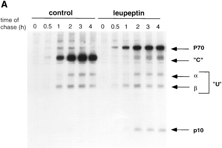
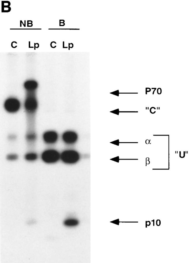
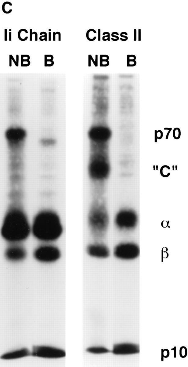
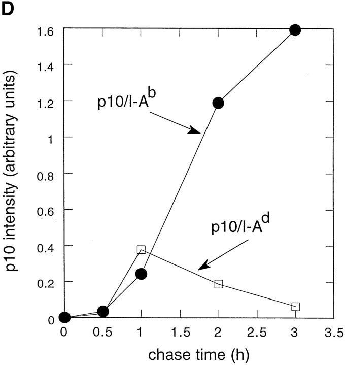
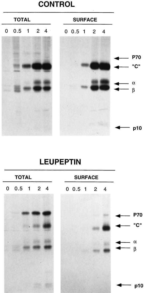
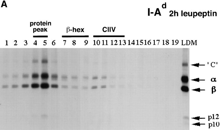
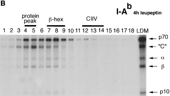
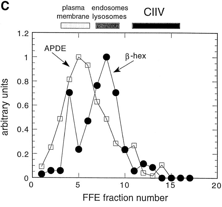
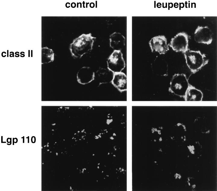
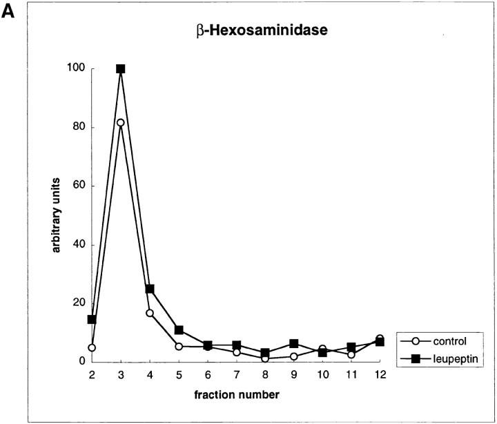
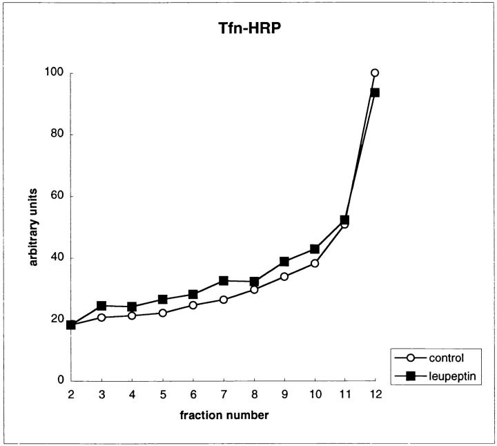
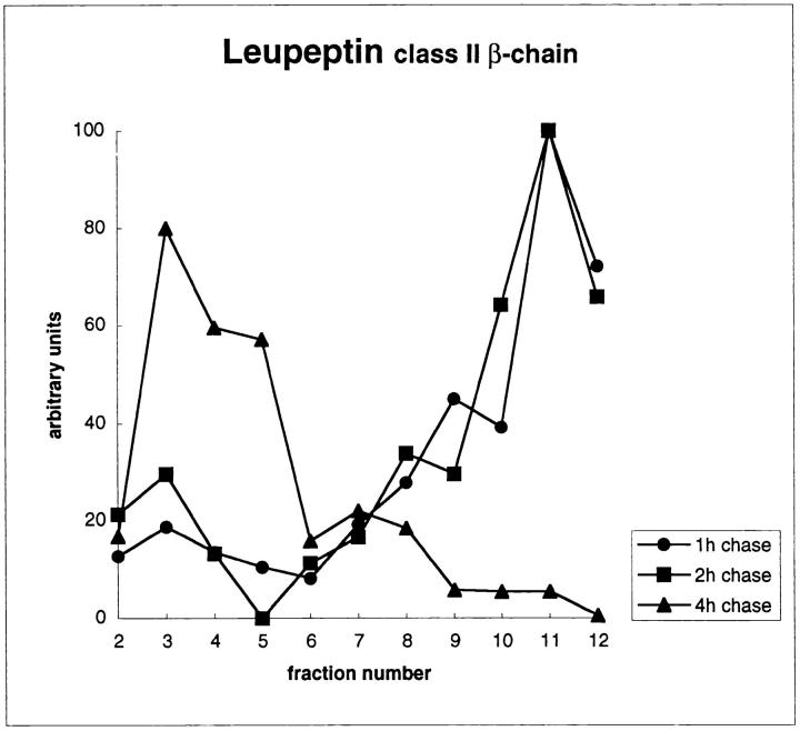
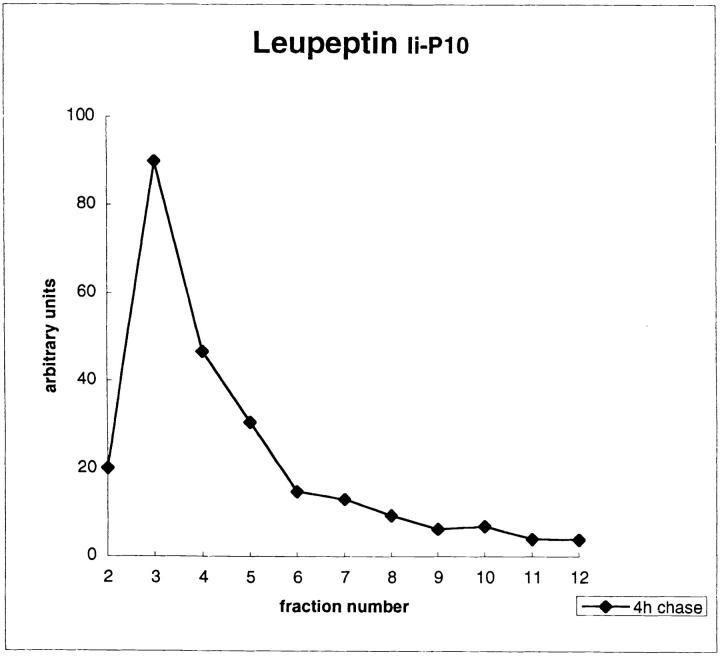
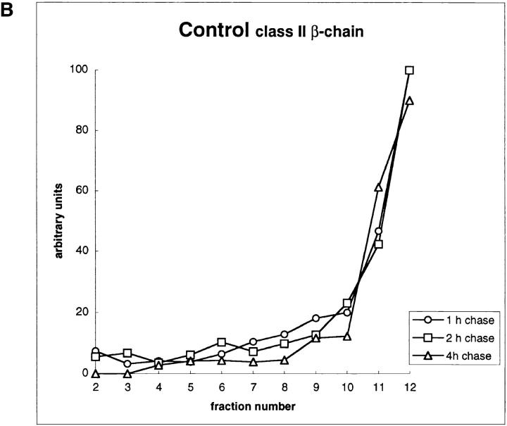
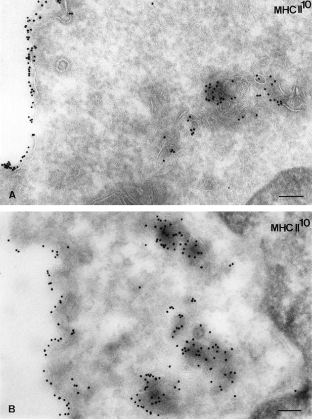
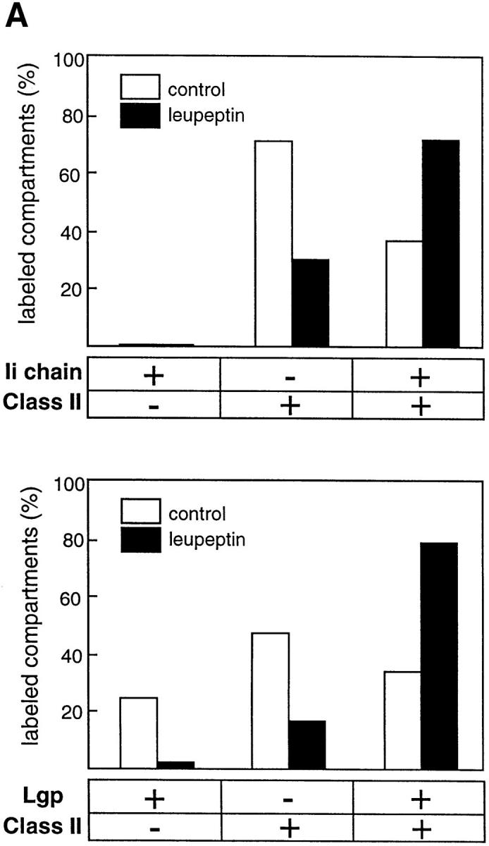
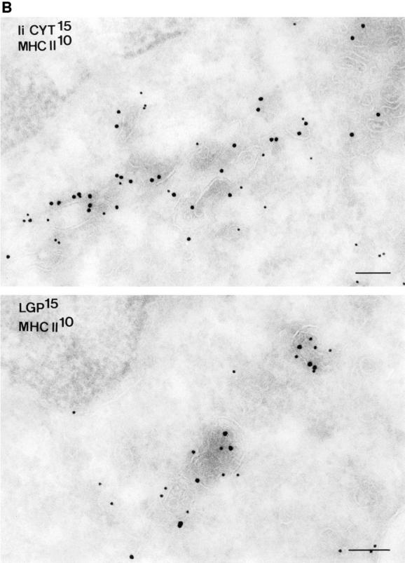
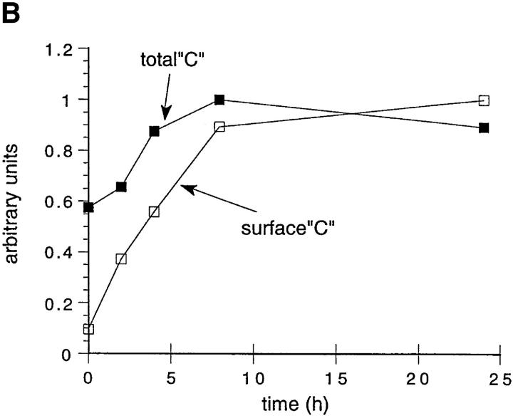
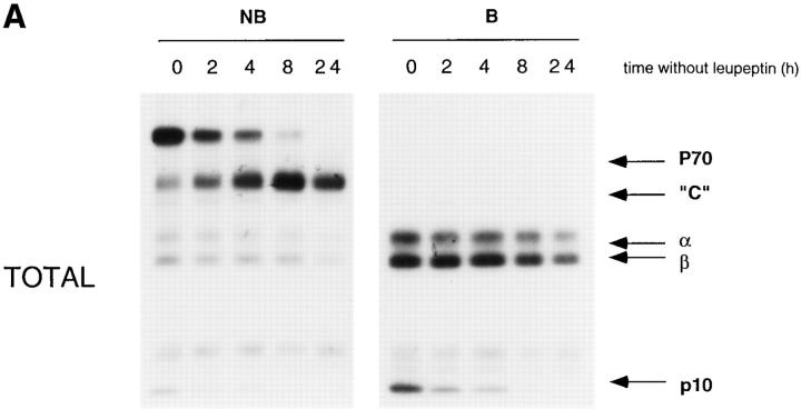
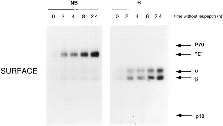
Major histocompatibility complex class II molecules are synthesized as a nonameric complex consisting of three alpha beta dimers associated with a trimer of invariant (Ii) chains. After exiting the TGN, a targeting signal in the Ii chain cytoplasmic domain directs the complex to endosomes where Ii chain is proteolytically processed and removed, allowing class II molecules to bind antigenic peptides before reaching the cell surface. Ii chain dissociation and peptide binding are thought to occur in one or more postendosomal sites related either to endosomes (designated CIIV) or to lysosomes (designated MIIC). We now find that in addition to initially targeting alpha beta dimers to endosomes, Ii chain regulates the subsequent transport of class II molecules. Under normal conditions, murine A20 B cells transport all of their newly synthesized class II I-A(b) alpha beta dimers to the plasma membrane with little if any reaching lysosomal compartments. Inhibition of Ii processing by the cysteine/serine protease inhibitor leupeptin, however, blocked transport to the cell surface and caused a dramatic but selective accumulation of I-A(b) class II molecules in lysosomes. In leupeptin, I-A(b) dimers formed stable complexes with a 10-kD NH2-terminal Ii chain fragment (Ii-p10), normally a transient intermediate in Ii chain processing. Upon removal of leupeptin, Ii-p10 was degraded and released, I-A(b) dimers bound antigenic peptides, and the peptide-loaded dimers were transported slowly from lysosomes to the plasma membrane. Our results suggest that alterations in the rate or efficiency of Ii chain processing can alter the postendosomal sorting of class II molecules, resulting in the increased accumulation of alpha beta dimers in lysosome-like MIIC. Thus, simple differences in Ii chain processing may account for the highly variable amounts of class II found in lysosomal compartments of different cell types or at different developmental stages.


Figures

Figure 1
Figure 1
Leupeptin induces an accumulation of SDS- resistant I-Ab Ii-p10 complexes. (A) Leupeptin induces the accumulation of 10-kD (Ii-p10) and 70-kD (p70) proteins that coprecipitate with I-Ab. I-Ab–expressing A20 cells were pulsed for 20 min with [35S]methionine and chased at 37°C for the indicated times in the presence or absence of 2 mM leupeptin. After lysis, the I-Ab molecules were immunoprecipitated using the Y3P mAb. The samples were not boiled before SDS-PAGE. Labeled class II molecules were not detected before 30 min of chase because the Y3P mAb used for immunoprecipitation does not detect immature αβ dimers complexed with intact Ii chain. (B) p70 represents SDS-stable complexes containing class II α and β chains and a 10-kD protein. After a 20-min pulse and 4-h chase with or without 2 mM leupeptin (lanes Lp and C, respectively), class II molecules were immunoprecipitated using the Y3P mAb, and the samples were boiled (B) or not boiled (NB) before SDSPAGE. After boiling, p70 dissociated quantitatively into monomers corresponding to αβ and Ii-p10. (C) p70 represents SDS-stable I-Ab αβ–Ii-p10 complexes. After a 20-min pulse and 4-h chase in the presence of leupeptin, class II molecules were immunoprecipitated with either anti–I-Ab (Y3P) or anti–Ii chain cytoplasmic domain (IN-1) mAbs. While both antibodies precipitated the p70 complex, only anti–class II mAb precipitated the 60-kD SDS-stable compact dimer. Thus, p70 but not compact dimers are complexed with Ii chain or Ii chain fragments (i.e., Ii-p10) that contain the Ii chain cytoplasmic domain. (D) Kinetics of association between Ii-p10 and I-Ab or I-Ad. Pulse-chase experiments were performed as above using A20 cells expressing only I-Ad or expressing both I-Ad and I-Ab. I-Ad or I-Ab–containing complexes were then immunoprecipitated using specific mAbs (Y3P and MKD6, respectively), and the amounts of Ii-p10 associated to the class II molecules were quantified by phosphorimaging. The association of Ii-p10 with I-Ab persisted throughout the chase period, while Ii-p10–I-Ad complexes appeared only transiently.
Figure 1
Figure 1
Leupeptin induces an accumulation of SDS- resistant I-Ab Ii-p10 complexes. (A) Leupeptin induces the accumulation of 10-kD (Ii-p10) and 70-kD (p70) proteins that coprecipitate with I-Ab. I-Ab–expressing A20 cells were pulsed for 20 min with [35S]methionine and chased at 37°C for the indicated times in the presence or absence of 2 mM leupeptin. After lysis, the I-Ab molecules were immunoprecipitated using the Y3P mAb. The samples were not boiled before SDS-PAGE. Labeled class II molecules were not detected before 30 min of chase because the Y3P mAb used for immunoprecipitation does not detect immature αβ dimers complexed with intact Ii chain. (B) p70 represents SDS-stable complexes containing class II α and β chains and a 10-kD protein. After a 20-min pulse and 4-h chase with or without 2 mM leupeptin (lanes Lp and C, respectively), class II molecules were immunoprecipitated using the Y3P mAb, and the samples were boiled (B) or not boiled (NB) before SDSPAGE. After boiling, p70 dissociated quantitatively into monomers corresponding to αβ and Ii-p10. (C) p70 represents SDS-stable I-Ab αβ–Ii-p10 complexes. After a 20-min pulse and 4-h chase in the presence of leupeptin, class II molecules were immunoprecipitated with either anti–I-Ab (Y3P) or anti–Ii chain cytoplasmic domain (IN-1) mAbs. While both antibodies precipitated the p70 complex, only anti–class II mAb precipitated the 60-kD SDS-stable compact dimer. Thus, p70 but not compact dimers are complexed with Ii chain or Ii chain fragments (i.e., Ii-p10) that contain the Ii chain cytoplasmic domain. (D) Kinetics of association between Ii-p10 and I-Ab or I-Ad. Pulse-chase experiments were performed as above using A20 cells expressing only I-Ad or expressing both I-Ad and I-Ab. I-Ad or I-Ab–containing complexes were then immunoprecipitated using specific mAbs (Y3P and MKD6, respectively), and the amounts of Ii-p10 associated to the class II molecules were quantified by phosphorimaging. The association of Ii-p10 with I-Ab persisted throughout the chase period, while Ii-p10–I-Ad complexes appeared only transiently.
Figure 2
Figure 2
Intracellular retention of Ii-p10–associated class II molecules. Cells were pulsed for 20 min and chased for the indicated times (h) in the presence or absence of 2 mM leupeptin. At each time point, the cells were surface biotinylated before lysis. Total and cell surface biotinylated class II molecules were sequentially immunoprecipitated using the mAb Y3P and streptavidin-agarose. The samples were then analyzed by SDS-PAGE without boiling before electrophoresis. In untreated control cells (top), peptideloaded, 60-kD compact αβ dimers (“C”) began to appear both in total lysates and on the surface after 0.5 h of chase. A small amount of the p70 complex of Ii-p10–αβ dimers appeared transiently, beginning also at 0.5 h of chase. In leupeptin-treated cells (bottom), p70 began to accumulate in total lysates by 0.5 h, but little was recovered at the plasma membrane. Peptide-loaded compact dimers only began to appear after 2–4 h of chase both in lysates and at the cell surface.
Figure 3
Figure 3
Fractionation of leupeptin-treated A20 cells by free flow electrophoresis. (A) A20 cells were pulse labeled for 20 min and chased for 2 h in the presence of leupeptin before fractionation by FFE. Membranes collected in each fraction were pelleted by centrifugation and lysed in Triton X-100, and I-Ab class II molecules were immunoprecipitated using mAb MKD6. The samples were then analyzed by SDS-PAGE without boiling. The positions of compact dimers (“C”), α, β chains, Ii-p10, and a p12 protein of unknown origin are indicated relative to the positions of markers for the major protein peak (plasma membrane), endosomes/lysosomes (β-hexosaminidase), and anodally shifted CIIVcontaining fractions. (Left) Anode; (right) cathode. (B) I-Ab– expressing A20 cells were pulse labeled for 20 min and chased for 4 h in the presence of leupeptin before fractionation by FFE. The positions of p70 (Ii-p10–αβ complexes), 60-kD peptide-loaded compact dimers, free α and β chains, and Ii-p10 are indicated. (C) Positions of marker enzymes for plasma membrane (alkaline phosphodiesterase) and endosomes/lysosomes (β-hexosaminidase) in the FFE profile shown in B. p70 codistributed largely with the lysosomal marker β-hexosaminidase.
Figure 3
Figure 3
Fractionation of leupeptin-treated A20 cells by free flow electrophoresis. (A) A20 cells were pulse labeled for 20 min and chased for 2 h in the presence of leupeptin before fractionation by FFE. Membranes collected in each fraction were pelleted by centrifugation and lysed in Triton X-100, and I-Ab class II molecules were immunoprecipitated using mAb MKD6. The samples were then analyzed by SDS-PAGE without boiling. The positions of compact dimers (“C”), α, β chains, Ii-p10, and a p12 protein of unknown origin are indicated relative to the positions of markers for the major protein peak (plasma membrane), endosomes/lysosomes (β-hexosaminidase), and anodally shifted CIIVcontaining fractions. (Left) Anode; (right) cathode. (B) I-Ab– expressing A20 cells were pulse labeled for 20 min and chased for 4 h in the presence of leupeptin before fractionation by FFE. The positions of p70 (Ii-p10–αβ complexes), 60-kD peptide-loaded compact dimers, free α and β chains, and Ii-p10 are indicated. (C) Positions of marker enzymes for plasma membrane (alkaline phosphodiesterase) and endosomes/lysosomes (β-hexosaminidase) in the FFE profile shown in B. p70 codistributed largely with the lysosomal marker β-hexosaminidase.
Figure 4
Figure 4
Leupeptin treatment causes MHC class II molecules to accumulate in lgp-containing structures by immunofluorescence microscopy. Control or leupeptintreated (3 h, 2 mM leupeptin) I-Ab–expressing A20 cells were fixed, permeabilized, and then stained for MHC class II (FITC, using mAb Y3P, two upper panels) vs lgp-B (TRITC, using mAb GL2A7, lower two panels). In control cells, the small amount of intracellular MHC class II was localized to structures that were generally negative for lgp-B. These probably represented CIIVs and early endosomes. Leupeptin treatment, however, induced extensive colocalization of class II and lgp-B.
Figure 5
Figure 5
Selective redistribution of newly synthesized MHC class II molecules to lysosomes in leupeptin-treated cells analyzed by Percoll gradient centrifugation. (A) Leupeptin does not affect the density of compartments containing β-hexosaminidase and Tfn-HRP, markers of lysosomes and endosomes, respectively. After a 3-h incubation with or without 2 mM leupeptin, cells were permitted to internalize Tfn coupled to HRP for 30 min at 37°C (in the presence or absence of leupeptin) before homogenization and fractionation on Percoll density gradients. The β-hexosaminidase and HRP activities were determined in individual fractions. High density fractions (bottom of the gradient) contained most of the β-hexosaminidase activity, while Tfn-HRP was mainly present in low density fractions. No difference was observed between leupeptin-treated and control cells. (B) Redistribution of newly synthesized MHC class II molecules into high density fractions in leupeptin-treated cells. After a pulse of [35S]methionine and a 1-, 2-, or 4-h chase in the absence (left) or presence (right) of leupeptin, I-Ab–expressing A20 cells were fractionated on Percoll gradients. The membranes in each of the gradient fractions were pelleted by centrifugation, lysed in Triton X-100, and then immunoprecipitated using mAbs to I-Ab (Y3P). The samples were analyzed by SDS-PAGE, and bands corresponding to class II β chains were quantified by phosphorimaging and optical densitometry. Ii-p10 intensity was also quantified after the 4-h chase in the presence of leupeptin (bottom left). Leupeptin caused a strong redistribution of class II into high density fractions. The majority of Ii-p10 was also found in lysosome-containing fractions. As expected, Ii-p10 was barely detectable in control cells and thus was not shown.
Figure 5
Figure 5
Selective redistribution of newly synthesized MHC class II molecules to lysosomes in leupeptin-treated cells analyzed by Percoll gradient centrifugation. (A) Leupeptin does not affect the density of compartments containing β-hexosaminidase and Tfn-HRP, markers of lysosomes and endosomes, respectively. After a 3-h incubation with or without 2 mM leupeptin, cells were permitted to internalize Tfn coupled to HRP for 30 min at 37°C (in the presence or absence of leupeptin) before homogenization and fractionation on Percoll density gradients. The β-hexosaminidase and HRP activities were determined in individual fractions. High density fractions (bottom of the gradient) contained most of the β-hexosaminidase activity, while Tfn-HRP was mainly present in low density fractions. No difference was observed between leupeptin-treated and control cells. (B) Redistribution of newly synthesized MHC class II molecules into high density fractions in leupeptin-treated cells. After a pulse of [35S]methionine and a 1-, 2-, or 4-h chase in the absence (left) or presence (right) of leupeptin, I-Ab–expressing A20 cells were fractionated on Percoll gradients. The membranes in each of the gradient fractions were pelleted by centrifugation, lysed in Triton X-100, and then immunoprecipitated using mAbs to I-Ab (Y3P). The samples were analyzed by SDS-PAGE, and bands corresponding to class II β chains were quantified by phosphorimaging and optical densitometry. Ii-p10 intensity was also quantified after the 4-h chase in the presence of leupeptin (bottom left). Leupeptin caused a strong redistribution of class II into high density fractions. The majority of Ii-p10 was also found in lysosome-containing fractions. As expected, Ii-p10 was barely detectable in control cells and thus was not shown.
Figure 6
Figure 6
Immunogold localization of MHC class II molecules in leupeptin-treated cells. Ultrathin cryosections were immunogold labeled with the anti–class II mAb M5.114 and protein A–gold (PAG-10). (A) In control cells, MHC class II molecules are found on the plasma membrane, in intracellular compartments characterized by the presence of internal membranes. (B) In leupeptin-treated cells (18-h treatment), MHC class II molecules were detected on the plasma membrane and accumulate in electron-dense compartments displaying internal membranes. Bars, 120 nm.
Figure 7
Figure 7
Distribution of MHC class II, Ii chain, and lgp-B in leupeptin-treated cells. (A) Quantification of MHC class II, Ii, and lgp-B in control and leupeptin-treated cells. The quantifications were carried out on ultrathin cryosections immunogold labeled for Ii chain (anti–Ii cytosolic tail mAb IN1) and MHC class II (rabbit anti–I-A cytoplasmic domain serum) or immunogold labeled for lgp-B (mAb GL2A7) and MHC class II (rabbit anti–I-A cytoplasmic domain serum) or immunogold labeled for lgp-B (mAb GL2A7) and MHC class II (rabbit anti–I-A cytoplasmic domain serum). In each case, 40 cell profiles were analyzed. (B) Immunogold localization of Ii chain, lgp-B, and I-A in leupeptin-treated cells. (Upper panel) Ultrathin cryosections were double immunogold labeled with the Ii chain antibody IN-1 (anti–Ii cytosolic domain; Ii CYT) and rabbit anti–I-A cytosolic tail polyclonal antibody. Ii chain accumulates in I-A–positive compartments displaying internal vesicles and electron-dense content. (Lower panel) Ultrathin cryosections were double immunogold labeled with the anti–lgp-B mAb GL2A7 and rabbit anti–I-A cytosolic tail polyclonal antibody. Class II molecules were visualized in electron-dense, lgp-B–positive compartments. The size of the gold particles is indicated. Bars, 120 nm.
Figure 7
Figure 7
Distribution of MHC class II, Ii chain, and lgp-B in leupeptin-treated cells. (A) Quantification of MHC class II, Ii, and lgp-B in control and leupeptin-treated cells. The quantifications were carried out on ultrathin cryosections immunogold labeled for Ii chain (anti–Ii cytosolic tail mAb IN1) and MHC class II (rabbit anti–I-A cytoplasmic domain serum) or immunogold labeled for lgp-B (mAb GL2A7) and MHC class II (rabbit anti–I-A cytoplasmic domain serum) or immunogold labeled for lgp-B (mAb GL2A7) and MHC class II (rabbit anti–I-A cytoplasmic domain serum). In each case, 40 cell profiles were analyzed. (B) Immunogold localization of Ii chain, lgp-B, and I-A in leupeptin-treated cells. (Upper panel) Ultrathin cryosections were double immunogold labeled with the Ii chain antibody IN-1 (anti–Ii cytosolic domain; Ii CYT) and rabbit anti–I-A cytosolic tail polyclonal antibody. Ii chain accumulates in I-A–positive compartments displaying internal vesicles and electron-dense content. (Lower panel) Ultrathin cryosections were double immunogold labeled with the anti–lgp-B mAb GL2A7 and rabbit anti–I-A cytosolic tail polyclonal antibody. Class II molecules were visualized in electron-dense, lgp-B–positive compartments. The size of the gold particles is indicated. Bars, 120 nm.
Figure 8
Figure 8
Reversal of the leupeptin block results in MHC class II transport from lysosomes to the cell surface. (A) Reversibility of the effect of leupeptin on the intracellular retention of I-Ab class II molecules. Cells were metabolically labeled for 20 min, and then chased in the continous presence of leupeptin for 4 h. Leupeptin was then removed, and the cells were incubated for 0–24 h. After cell surface biotinylation, the cells were lysed, and total (left) or surface (right) I-Ab molecules were immunoprecipitated sequentially. Upon removal of leupeptin, the total amount of p70 (the Ii-p10–αβ complex) slowly decreased, while the amount of peptide-loaded, SDS-stable compact dimers (“C”) increased. Ii-p10 completely disappeared over this time course. After a lag (see B), compact dimers began to appear at the cell surface. (B) Kinetics of compact dimer formation and transport to the cell surface. Bands corresponding to total and surface compact dimers were quantified by phosphorimaging. The lag between formation of compact dimers and their subsequent appearance at the cell surface was 3–4 h.
Figure 8
Figure 8
Reversal of the leupeptin block results in MHC class II transport from lysosomes to the cell surface. (A) Reversibility of the effect of leupeptin on the intracellular retention of I-Ab class II molecules. Cells were metabolically labeled for 20 min, and then chased in the continous presence of leupeptin for 4 h. Leupeptin was then removed, and the cells were incubated for 0–24 h. After cell surface biotinylation, the cells were lysed, and total (left) or surface (right) I-Ab molecules were immunoprecipitated sequentially. Upon removal of leupeptin, the total amount of p70 (the Ii-p10–αβ complex) slowly decreased, while the amount of peptide-loaded, SDS-stable compact dimers (“C”) increased. Ii-p10 completely disappeared over this time course. After a lag (see B), compact dimers began to appear at the cell surface. (B) Kinetics of compact dimer formation and transport to the cell surface. Bands corresponding to total and surface compact dimers were quantified by phosphorimaging. The lag between formation of compact dimers and their subsequent appearance at the cell surface was 3–4 h.

References

    1. Amigorena S, Drake JR, Webster P, Mellman I. Transient accumulation of new class II molecules in a novel endocytic compartment in B lymphocytes. Nature (Lond) 1994;369:113–120. - PubMed
    1. Amigorena S, Webster P, Drake J, Newcomb J, Cresswell P, Mellman I. Invariant chain cleavage and peptide loading in major histocompatibility complex class II vesicles. J Exp Med. 1995;181:1729–1741. - PMC - PubMed
    1. Arneson LS, Miller J. Efficient endosomal localization of major histocompatibility complex class II–invariant chain complexes requires multimerization of the invariant chain targeting sequence. J Cell Biol. 1995;129:1217–1228. - PMC - PubMed
    1. Bakke O, Dobberstein B. MHC class II-associated invariant chain contains a sorting signal for endosomal compartments. Cell. 1990;63:707–716. - PubMed
    1. Barnes KA, Mitchell RN. Detection of functional class II-associated antigen: role of a low density endosomal compartment in antigen processing. J Exp Med. 1995;181:1715–1727. - PMC - PubMed

Publication types

MeSH terms