. 1997 Apr 7;137(1):93-104.
doi: 10.1083/jcb.137.1.93.

Lysosomes behave as Ca2+-regulated exocytic vesicles in fibroblasts and epithelial cells

Affiliations

Lysosomes behave as Ca2+-regulated exocytic vesicles in fibroblasts and epithelial cells

A Rodríguez et al. J Cell Biol. .

Abstract

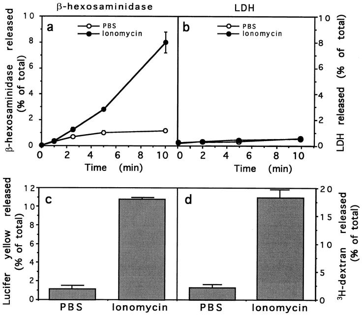
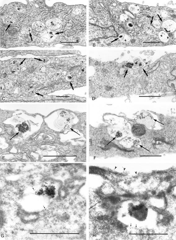
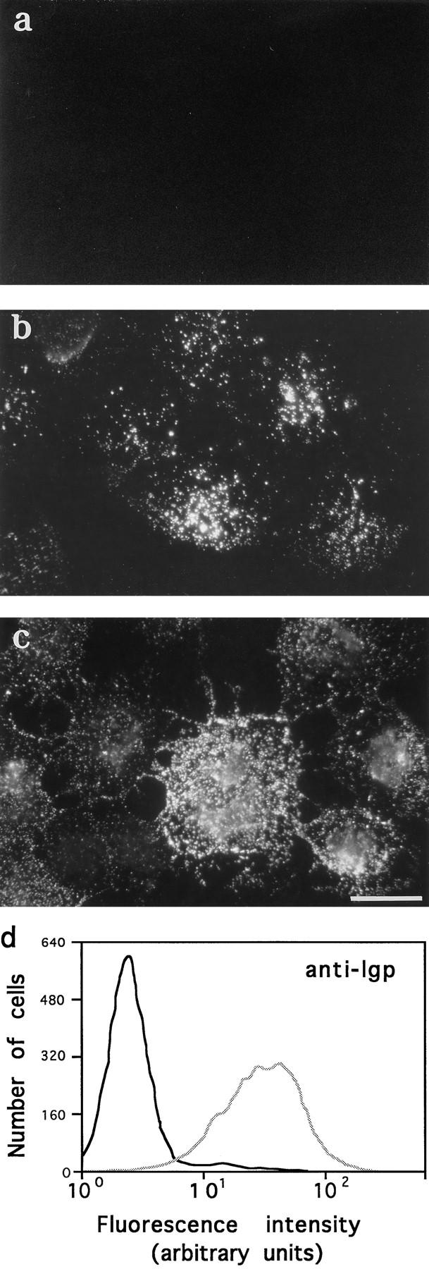
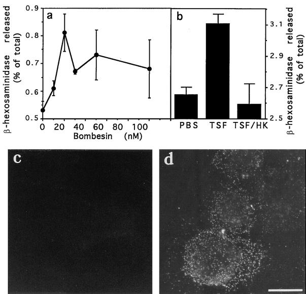
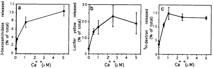
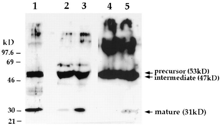
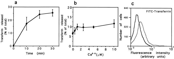
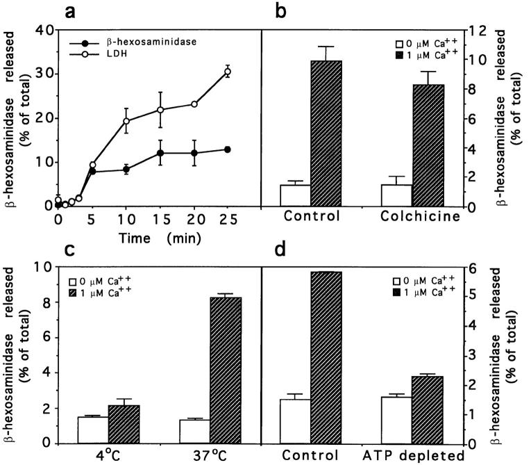
Lysosomes are considered to be a terminal degradative compartment of the endocytic pathway, into which transport is mostly unidirectional. However, specialized secretory vesicles regulated by Ca2+, such as neutrophil azurophil granules, mast cell-specific granules, and cytotoxic lymphocyte lytic granules, share characteristics with lysosomes that may reflect a common biogenesis. In addition, the involvement of Ca2+ transients in the invasion mechanism of the parasite Trypanosoma cruzi, which occurs by fusion of lysosomes with the plasma membrane, suggested that lysosome exocytosis might be a generalized process present in most cell types. Here we demonstrate that elevation in the intracellular free Ca2+ concentration of normal rat kidney (NRK) fibroblasts induces fusion of lysosomes with the plasma membrane. This was verified by measuring the release of the lysosomal enzyme beta-hexosaminidase, the appearance on the plasma membrane of the lysosomal glycoprotein lgp120, the release of fluid-phase tracers previously loaded into lysosomes, and the release of the lysosomally processed form of cathepsin D. Exposure to the Ca2+ ionophore ionomycin or addition of Ca2+-containing buffers to streptolysin O-permeabilized cells induced exocytosis of approximately 10% of the total lysosomes of NRK cells. The process was also detected in other cell types such as epithelial cells and myoblasts. Lysosomal exocytosis was found to require micromolar levels of Ca2+ and to be temperature and ATP dependent, similar to Ca2+-regulated secretory mechanisms in specialized cells. These findings highlight a novel role for lysosomes in cellular membrane traffic and suggest that fusion of lysosomes with the plasma membrane may be an ubiquitous form of Ca2+-regulated exocytosis.


Figures

Figure 1
Figure 1
Ionomycin induces exocytosis of β-hexosaminidase and lysosomal fluid-phase tracers but not LDH from intact NRK fibroblasts. (a and b) NRK cells were incubated at 37°C with either PBS (open circles) or 10 μM ionomycin in PBS (black circles). At the indicated time points, the incubation buffer was collected and assayed for β-hexosaminidase and LDH activity. The amount of enzyme released at each point is expressed as a percentage of the total content of enzyme in control cells. (c and d) Lysosomes of NRK cells were loaded with lucifer yellow or 3H-dextran by fluidphase endocytosis followed by a 2-h chase, before treatment with PBS or 10 μM ionomycin for 10 min. The incubation buffer was collected, lucifer yellow was detected by reading the fluorescence, and 3H-dextran was detected by scintillation counting. The amount of lucifer yellow or 3H-dextran released in each sample is expressed as a percentage of the total amount of tracer present in control cells. The data represent the average of triplicate determinations ±SD.
Figure 2
Figure 2
Exocytosis of BSA–gold-loaded lysosomes is triggered by [Ca2+]i elevation. NRK cells loaded with 5 nm BSA–gold complexes for 4 h followed by a chase of 2 h were incubated with PBS (A and B) or 10 μM ionomycin (C–H) for 2.5 (C and D) or 10 min (E–H). Transmission EM sections show gold-loaded vesicles (arrows), observed in close proximity to the plasma membrane in ionomycintreated cells (C, E, and F). Exocytosed BSA–gold was also detected after exposure to ionomycin (D and F). Labeling with antibodies against lgp120 (10 nm gold) was detected on BSA–gold-containing vesicles (G and H). Small arrows indicate anti-lgp120 labeling on lysosomes; arrowheads indicate anti-lgp120 labeling on the plasma membrane after ionomycin treatment. Bars, 1 μM.
Figure 3
Figure 3
Ionomycin induces appearance of lgp120 on the plasma membrane. NRK cells, either attached to coverslips (a–c) or in suspension (d), were incubated with PBS or 10 μM ionomycin in PBS at 37°C, followed by immunofluorescent surface labeling with an mAb to a luminal domain of lgp 120. (a) lgp120 surface staining 10 min after exposure to PBS; (b) lgp120 surface staining 3 min after exposure to ionomycin; (c) lgp120 surface staining 10 min after exposure to ionomycin. (d) FACS® analysis of NRK cells in suspension treated for 5 min with PBS (black line) or 10 μM ionomycin in PBS (gray line). Cells were incubated with antilgp120 mAbs for 30 min at 4°C before fixation. Bar, 5 μm.
Figure 4
Figure 4
Ca2+ agonists induce a low level of β-hexosaminidase release and appearance of lgp120 in the plasma membrane. (a) NRK cells were incubated with different concentrations of bombesin for 5 min, and the incubation buffer was assayed for β-hexosaminidase activity. The amount of enzyme released at each point is expressed as a percentage of the total content of enzyme in control cells. The data represent the average of triplicate determinations ± SD. (b) Same as a, except that cells were incubated for 5 min with PBS, TSF, or heat-inactivated TSF. (c) lgp120 surface staining 5 min after exposure to PBS; (d) lgp120 surface staining of NRK cells 5 min after exposure to 10 μM bombesin. Bar, 5 μM.
Figure 5
Figure 5
Release of β-hexosaminidase and LDH from intact and SLO-permeabilized NRK cells. Intact cells (black columns) and SLO-permeabilized cells (white columns), were incubated in permeabilization buffer containing either 0, 1, or 5 μM Ca2+ for 5 min at 37°C. Supernatants were assayed for (a) β-hexosaminidase and (b) LDH activity. The amount of enzyme released in each sample is expressed as a percentage of the total enzyme content of control cells. The data represent the average of triplicate determinations ±SD.
Figure 6
Figure 6
Ca2+-dependent release of β-hexosaminidase, lucifer yellow, and 3H-dextran from permeabilized NRK cells. SLO-permeabilized cells were incubated for 5 min at 37°C in permeabilization buffer containing different Ca2+ concentrations. (a) β-hexosaminidase activity released. (b) Lucifer yellow released from previously loaded cells. (c) 3H-Dextran released from previously loaded cells. Values are expressed as a percentage of the total content of either β-hexosaminidase, lucifer yellow, or 3H-dextran in control cells. The data represent the average of triplicate determinations ±SD.
Figure 7
Figure 7
Elevation in [Ca2+]i induces secretion of the 31-kD lysosomally processed form of cathepsin D. Rabbit anti–cathepsin D antibodies were used to probe a Western blot containing a lysate and concentrated supernatants of IMR-90 human fibroblasts. (Lane 1) total lysate; (lane 2) concentrated supernatant of cells treated with PBS for 5 min; (lane 3) concentrated supernatant of cells treated with 10 μM ionomycin for 5 min; (lanes 4 and 5) concentrated supernatants of SLO-permeabilized cells incubated for 5 min in buffers containing 0 or 1 μM Ca2+, respectively. Different ECL exposures were performed for each treatment to allow visualization of the secreted mature 31-kD cathepsin D band.
Figure 8
Figure 8
Effect of elevated [Ca2+]i on exocytosis of transferrin receptor–containing endosomes. (a) Kinetics of release of preinternalized 125I-transferrin from SLOpermeabilized NRK cells in the absence of Ca2+. (b) Release of preinternalized 125Itransferrin from SLO-permeabilized NRK cells at different Ca2+ concentrations. Values are expressed as a percentage of the total 125Itransferrin present in each sample (cell associated plus released). The data represent the average of triplicate determinations ±SD. (c) FACS® analysis of NRK cells in suspension treated for 5 min with PBS (black line) or 10 mM ionomycin in PBS (gray line). Cells were incubated with FITC-transferrin for 30 min at 4°C before fixation.
Figure 9
Figure 9
Characterization of Ca2+-induced lysosome exocytosis in permeabilized cells. Release of β-hexosaminidase from SLOpermeabilized NRK cells was measured after addition of 0 or 1 μM Ca2+ buffers. (a) Kinetics of release of β-hexosaminidase (black circles) and LDH (white circles) from NRK cells after addition of 1 μM Ca2+ at 37°C. (b) Sensitivity of β-hexosaminidase release to microtubule depolymerization. Cells were treated with 10 μM colchicine for 1 h before addition of the Ca2+ buffers for 5 min at 37°C. (c) Temperature dependence of β-hexosaminidase release. Cells were incubated for 5 min at 37°C to allow SLO pore formation, and then the secretion assay was performed at 4° or 37°C for 5 min. (d) Sensitivity of β-hexosaminidase release to ATP depletion. After permeabilization, cells were incubated in the presence of 2 mM MgATP or an ATP-depleting system (5 mM glucose, 150 U/ml hexokinase) for 15 min at 37°C, before performing the 5-min secretion assay. In all experiments, the amount of enzyme released is expressed as a percentage of the total enzyme content of control cells. The data represent the average of triplicate determinations ±SD.

References

    1. Adams CJ, Maurey KM, Storrie B. Exocytosis of pinocytic contents by Chinese hamster ovary cells. J Cell Biol. 1982;93:632–637. - PMC - PubMed
    1. Ahnert-Hilger G, Mach W, Fohr KJ, Gratzl M. Poration by α-toxin and streptolysin-O: an approach to analyze intracellular processes. Methods Cell Biol. 1989;31:63–90. - PubMed
    1. Baron R, Neff L, Brown W, Louvard D, Courtnoy PJ. Selective internalization of the apical plasma membrane and rapid redistribution of lysosomal enzymes and mannose-6-phosphate receptors during osteoclast inactivation by calcitonin. J Cell Sci. 1990;97:439–447. - PubMed
    1. Beckers CJM, Balch WE. Calcium and GTP: essential components in vesicular trafficking between the endoplasmic reticulum and Golgi apparatus. J Cell Biol. 1989;108:1245–1256. - PMC - PubMed
    1. Bi G-Q, Alderton JM, Steinhardt RA. Calcium-regulated exocytosis is required for cell membrane resealing. J Cell Biol. 1995;131:1747–1758. - PMC - PubMed

Publication types

MeSH terms