. 1997 Apr 7;137(1):131-40.
doi: 10.1083/jcb.137.1.131.

Point mutations in human beta cardiac myosin heavy chain have differential effects on sarcomeric structure and assembly: an ATP binding site change disrupts both thick and thin filaments, whereas hypertrophic cardiomyopathy mutations display normal assembly

Affiliations

Point mutations in human beta cardiac myosin heavy chain have differential effects on sarcomeric structure and assembly: an ATP binding site change disrupts both thick and thin filaments, whereas hypertrophic cardiomyopathy mutations display normal assembly

K D Becker et al. J Cell Biol. .

Abstract

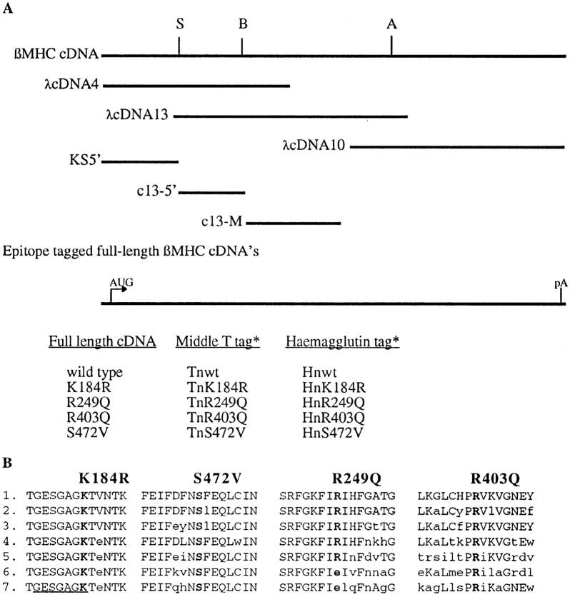
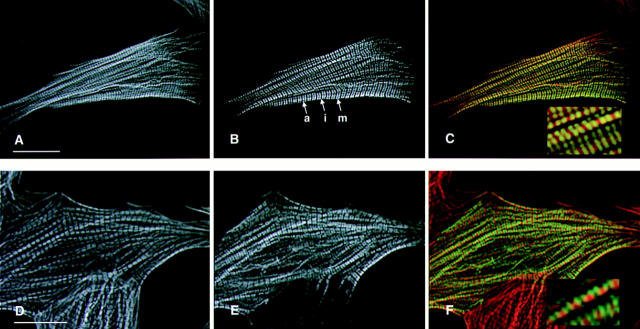
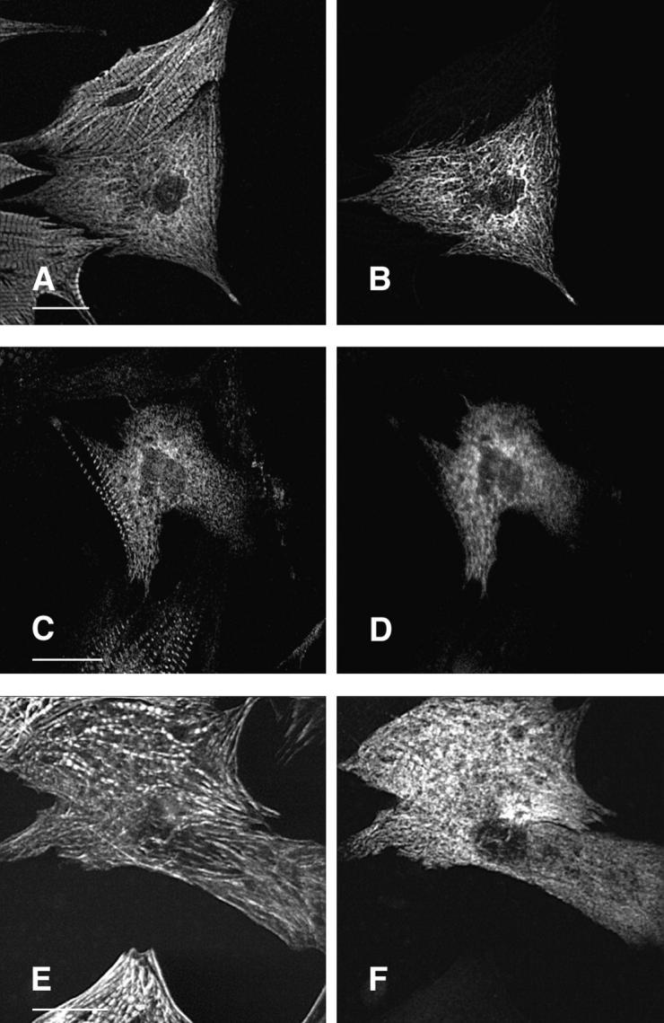
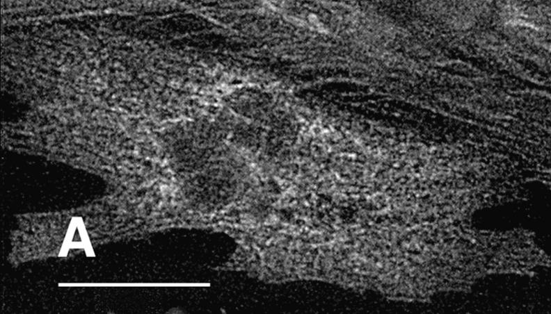
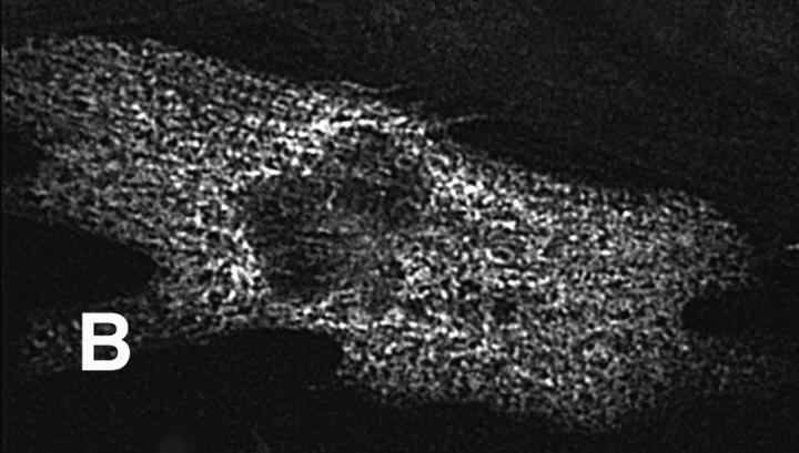
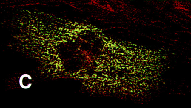
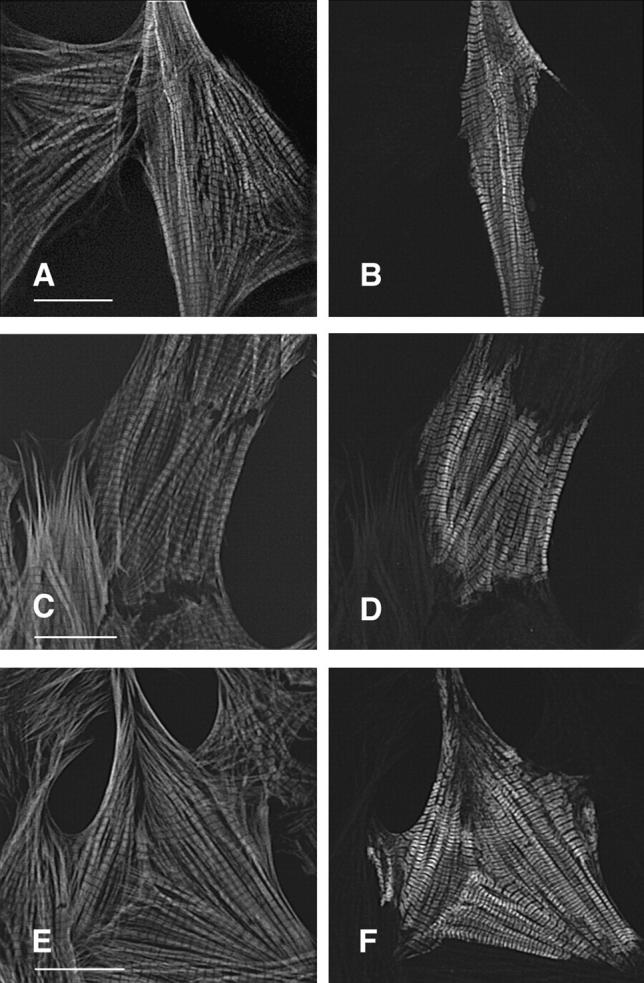
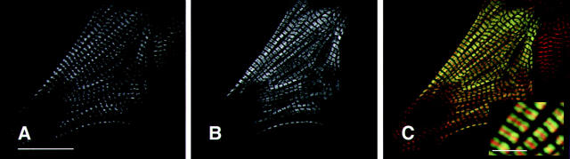
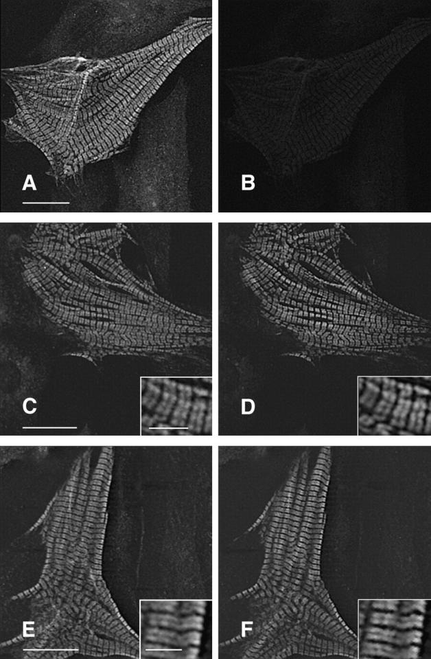
Hypertrophic cardiomyopathy is a human heart disease characterized by increased ventricular mass, focal areas of fibrosis, myocyte, and myofibrillar disorganization. This genetically dominant disease can be caused by mutations in any one of several contractile proteins, including beta cardiac myosin heavy chain (beta MHC). To determine whether point mutations in human beta MHC have direct effects on interfering with filament assembly and sarcomeric structure, full-length wild-type and mutant human beta MHC cDNAs were cloned and expressed in primary cultures of neonatal rat ventricular cardiomyocytes (NRC) under conditions that promote myofibrillogenesis. A lysine to arginine change at amino acid 184 in the consensus ATP binding sequence of human beta MHC resulted in abnormal subcellular localization and disrupted both thick and thin filament structure in transfected NRC. Diffuse beta MHC K184R protein appeared to colocalize with actin throughout the myocyte, suggesting a tight interaction of these two proteins. Human beta MHC with S472V mutation assembled normally into thick filaments and did not affect sarcomeric structure. Two mutant myosins previously described as causing human hypertrophic cardiomyopathy, R249Q and R403Q, were competent to assemble into thick filaments producing myofibrils with well defined I bands, A bands, and H zones. Coexpression and detection of wild-type beta MHC and either R249Q or R403Q proteins in the same myocyte showed these proteins are equally able to assemble into the sarcomere and provided no discernible differences in subcellular localization. Thus, human beta MHC R249Q and R403Q mutant proteins were readily incorporated into NRC sarcomeres and did not disrupt myofilament formation. This study indicates that the phenotype of myofibrillar disarray seen in HCM patients which harbor either of these two mutations may not be directly due to the failure of the mutant myosin heavy chain protein to assemble and form normal sarcomeres, but may rather be a secondary effect possibly resulting from the chronic stress of decreased beta MHC function.


Figures

Figure 1
Figure 1
Cloning and construction of human βMHC cDNA expression plasmids. (A) The three overlapping cDNA clones (λcDNA4, 13, and 10) used for construction of the 6-kbp, fulllength human βMHC cDNA are shown below the representation of the full-length cDNA (βMHC cDNA). The restriction sites SmaI (S), BamHI (B), and AatII (A) were used to assemble the full-length clone. Subclones KS5′, c13-5′, and c13-M were used to insert the epitope tag sequences or for generation of the mutant codons. Also listed are the full-length, epitope tagged clones used. (B) Single letter amino acid sequence of wild-type myosin from several species are compared. The numbering is based upon the published human βMHC sequence (Liew et al., 1990). The underlined area is involved in ATP binding. 1, human βMHC; 2, chick sarcomeric MHC (Molina et al., 1987); 3, rat embryonal sarcomeric MHC (Strehler et al., 1986); 4, Ceanorhabditis elegans MHC A (Karn et al., 1983); 5, chick smooth muscle MHC (Yanagisawa et al., 1987); 6, Dictyostelium discoideum (Warrick et al., 1986); 7, Acathamoeba castellanii myosin II (Hammer et al., 1987).
Figure 2
Figure 2
Epitope tagged wild-type human βMHC protein assembles into normal myofibrils in transiently transfected NRC. Cells were transfected with plasmids containing the wild-type human βMHC cDNA tagged with either the EE (A–C) or HA (D–F). The cells were stained for the presence of actin filaments using rhodamine–phalloidin (A and C) and were costained with the anti-EE (B) or anti-HA (E) specific antibodies. Sarcomeres containing human βMHC have well defined A bands (a), I bands (i), and M lines (m). The composite images (C and F) show that the exogenous βMHC (green) fills the A band, except the H zone as expected, in a pattern that is complementary and partially overlapping with F-actin (red, C and F). Bar, 20 μm (inset enlarged 3×).
Figure 3
Figure 3
Expression of K184R βMHC in NRC disrupts myofibril assembly. NRC transfected with the human βMHC K184R plasmid were stained for with the anti-EE epitope specific antibody (B, D, and F). These cells were costained with anti-βMHC (A), anti-myomesin (C), or rhodamine–phalloidin (E). Sequential staining with two primary monoclonal antibodies is described in Materials and Methods. Bar, 20 μm.
Figure 4
Figure 4
K184R protein colocalizes with actin. A transfected NRC expressing K184R was costained with monoclonal antibody A20 (anti-actin, A) and the epitope specific antibody (anti-EE, B). The composite image (C) shows the high degree of colocalization of these two proteins throughout the cell. Bar, 20 μm.
Figure 4
Figure 4
K184R protein colocalizes with actin. A transfected NRC expressing K184R was costained with monoclonal antibody A20 (anti-actin, A) and the epitope specific antibody (anti-EE, B). The composite image (C) shows the high degree of colocalization of these two proteins throughout the cell. Bar, 20 μm.
Figure 5
Figure 5
S472V as well as hypertrophic cardiomyopathy mutants R249Q and R403Q assemble normally and do not disrupt myofibril structure. Cells transfected with R249Q (A and B), R403Q (C and D), or S472V (E and F) were stained for the presence of exogenous myosin with the epitope specific antibody (B, D, and F) and with rhodamine–phalloidin (A, C, and E). Bar, 20 μm.
Figure 6
Figure 6
Thick filaments containing R403Q have normal organization within the sarcomere. NRC transfected with human R403Q plasmid were costained for exogenous βMHC (B) with the epitope specific antibody and myomesin (A). The merged image (C) shows that myomesin staining is completely localized to the M-line, indicating normal thick filament and sarcomere structure. Bars: (A) 20 μm; (inset), 5 μm.
Figure 7
Figure 7
Coexpression of both mutant and wild-type βMHC molecules detects no differences in subcellular localization of the two exogenous proteins. NRC were cotransfected with both wildtype and mutant expression plasmids, each tagged with a different epitope. Sequential staining (see Materials and Methods) allows detection of each protein within the same cell. A and B show a cardiomyocyte transfected with Hnwt only. This cell was immunostained with anti-HA, goat anti–mouse–FITC (F(ab)), and anti- EE and then donkey anti–mouse–LRSC. A shows the anti-HA specific staining (FITC channel), and B shows the LRSC channel, indicating that the anti-EE antibody does not crossreact, and minimal signal bleed through occurs under these conditions. Using anti-EE to detect Tnwt, the same lack of crossreactivity is observed when costaining with anti-HA (data not shown). Expression and detection of both Hnwt (C) and Tnwt (D) in the same cell shows identical subcellular localization for both proteins. The TnR403Q mutant (F) also shows identical distribution to Hnwt βMHC (E) when coexpressed in the same cell. Similar results are obtained when coexpressing either R249Q or S472V with wildtype βMHC. The type of epitope does not affect the outcome of this experiment. Bars: (A, C, and E) 20 μm; (E and inset) 5 μm.

References

    1. Beall CJ, Sepanski MA, Fyrberg EA. Genetic dissection of Drosophilamyofibril formation: effects of actin and myosin heavy chain null alleles. Genes Dev. 1989;3:131–140. - PubMed
    1. Bejsovec A, Anderson P. Myosin heavy-chain mutations that disrupt Caenorhabditis elegansthick filament assembly. Genes Dev. 1988;2:1307–1317. - PubMed
    1. Bejsovec A, Anderson P. Functions of the myosin ATP and actin binding sites are required for C. elegansthick filament assembly. Cell. 1990;60:133–140. - PubMed
    1. Bonne G, Carrier L, Bercovici J, Cruaud C, Richard P, Hainque B, Gautel M, Labeit S, James M, Beckmann J, et al. Cardiac myosin binding protein-C gene splice acceptor site mutation is associated with familial hypertrophic cardiomyopathy. Nat Genet. 1995;11:438–440. - PubMed
    1. Chen CA, Okayama H. Calcium phosphate-mediated gene transfer: a highly efficient transfection system for stably transforming cells with plasmid DNA. Biotech. 1988;6:632–638. - PubMed

Publication types

MeSH terms