. 1997 Apr 7;137(1):141-53.
doi: 10.1083/jcb.137.1.141.

The yeast gene, MDM20, is necessary for mitochondrial inheritance and organization of the actin cytoskeleton

Affiliations

The yeast gene, MDM20, is necessary for mitochondrial inheritance and organization of the actin cytoskeleton

G J Hermann et al. J Cell Biol. .

Abstract

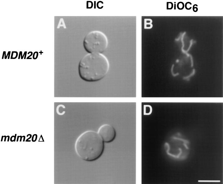
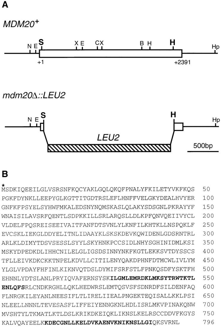
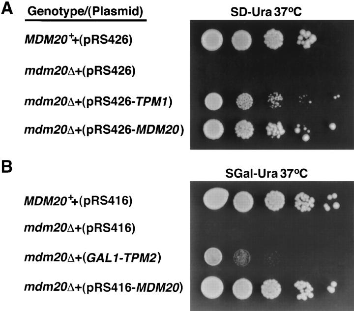
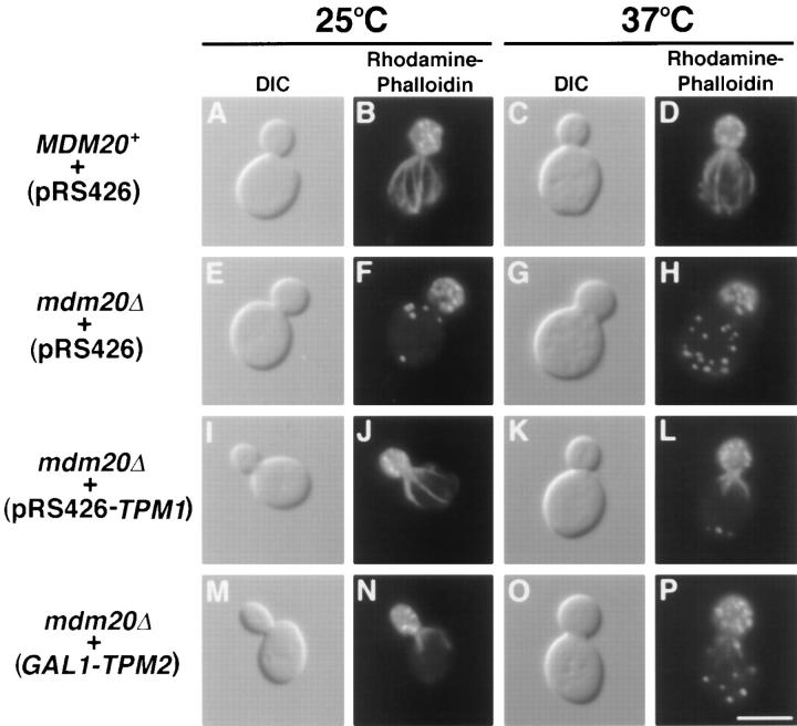
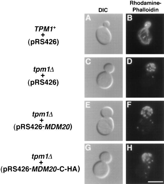
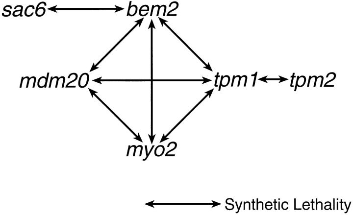
In Saccharomyces cerevisiae, the growing bud inherits a portion of the mitochondrial network from the mother cell soon after it emerges. Although this polarized transport of mitochondria is thought to require functions of the cytoskeleton, there are conflicting reports concerning the nature of the cytoskeletal element involved. Here we report the isolation of a yeast mutant, mdm20, in which both mitochondrial inheritance and actin cables (bundles of actin filaments) are disrupted. The MDM20 gene encodes a 93-kD polypeptide with no homology to other characterized proteins. Extra copies of TPM1, a gene encoding the actin filament-binding protein tropomyosin, suppress mitochondrial inheritance defects and partially restore actin cables in mdm20 delta cells. Synthetic lethality is also observed between mdm20 and tpm1 mutant strains. Overexpression of a second yeast tropomyosin, Tpm2p, rescues mutant phenotypes in the mdm20 strain to a lesser extent. Together, these results provide compelling evidence that mitochondrial inheritance in yeast is an actin-mediated process. MDM20 and TPM1 also exhibit the same pattern of genetic interactions; mutations in MDM20 are synthetically lethal with mutations in BEM2 and MYO2 but not SAC6. Although MDM20 and TPM1 are both required for the formation and/or stabilization of actin cables, mutations in these genes disrupt mitochondrial inheritance and nuclear segregation to different extents. Thus, Mdm20p and Tpm1p may act in vivo to establish molecular and functional heterogeneity of the actin cytoskeleton.


Figures

Figure 1
Figure 1
Mitochondrial inheritance during mitotic cell division. Mitochondria are localized near the cortex of unbudded and budding cells. Early in the cell cycle (S phase) a portion of the maternal mitochondrial network extends into the developing bud. As the bud grows (S phase to G2 phase), mitochondria continue to accumulate in the daughter cell. At cytokinesis (M phase/G1 phase boundary), the mitochondrial content of the daughter cell is often greater than that of the mother cell.
Figure 2
Figure 2
Cells lacking MDM20 have normal mitochondrial morphology but do not segregate mitochondria into buds at 37°C. DIC images (A and C) and DiOC6 mitochondrial staining (B and D) of MDM20 + (JSY999) (A and B) and mdm20Δ (JSY1065) (C and D) cells grown at 37°C for 3 h. Bar, 5 μm.
Figure 3
Figure 3
MDM20 encodes a novel 93-kD protein. (A, top) Restriction map of the MDM20 gene (open box). (A, bottom) The LEU2 construct used to generate the MDM20 disruption that replaces 2,219 bp (codons 12–751) of the MDM20 coding region. All restriction enzyme sites in the MDM20 gene are indicated: B, BglII; C, ClaI; E, EcoRI; H, HindIII; Hp, HpaI; S, SpeI; and X, XbaI. (B) Predicted amino acid sequence (single letter code) of the Mdm20p. The sequence begins at the first potential initiating methionine (asterisk). Indicated in boldface are two putative heptad repeats predicted by the COILS 2.1 program (Lupas et al., 1991). These sequence data are available from GenBank/EMBL/ DDBJ under accession number U54799.
Figure 4
Figure 4
Extra copies of TPM1 and TPM2 can suppress the temperature-sensitive growth defect in mdm20Δ cells. (A) MDM20 + (JSY999) or isogenic mdm20Δ (JSY1065) strains harboring pRS426, pRS426-MDM20, or pRS426-TPM1 plasmids were serially diluted and spotted on SD minus uracil (SDUra) solid medium and grown at 37°C for 3 d. (B) MDM20 + (JSY999) or isogenic mdm20Δ (JSY1065) strains harboring pRS416, pRS416- MDM20, or GAL1-TPM2 plasmids were serially diluted and spotted on SGal minus uracil (SGalUra) solid medium and grown at 37°C for 3 d.
Figure 5
Figure 5
Actin cables lacking in mdm20Δ cells are partially restored by introducing extra copies of TPM1 and TPM2. MDM20 + cells (JSY999) carrying pRS426 (A–D) and mdm20Δ cells (JSY1065) carrying pRS426 (E–H), pRS426-TPM1 (I–L), or GAL1-TPM2 (M–P) were grown in SD − Ura or SGal − Ura at 25°C, and then shifted to 25° and 37°C for 3 h. Cells were stained with rhodamine-phalloidin and visualized by DIC or fluorescence microscopy as indicated. Representative cells are shown. Bar, 5 μm.
Figure 6
Figure 6
Actin cables lacking in tpm1Δ cells are not restored by overexpression of Mdm20p. TPM1 + cells (FY22) containing pRS426 (A and B) and tpm1Δ cells (JSY707) containing pRS426 (C and D), pRS426-MDM20 (E and F), or pRS426-MDM20-CHA (G and H) were grown at 25°C, stained with rhodamine-phalloidin, and visualized by DIC or fluorescence microscopy as indicated. Representative cells are shown. Bar, 5 μm.
Figure 7
Figure 7
Expression of Mdm20p-C-HA in wild-type and tpm1Δ cells. Extracts were prepared from wild-type cells (JSY999) containing pRS416 (lane 1), pRS426 (lane 2), pRS416-MDM20-CHA (lane 3), and pRS426-MDM20-C-HA (lane 4), or tpm1Δ cells (JSY707) containing pRS416 (lane 5), pRS426 (lane 6), pRS416- MDM20-C-HA (lane 7), and pRS426-MDM20-C-HA (lane 8). The cells were harvested after growth to mid log phase at 25°C, and equal amounts (equivalent OD units) of the cell extracts were subjected to SDS-PAGE and transferred to nitrocellulose. The nitrocellulose was probed with antibodies to the HA epitope. The arrowhead identifies the Mdm20-C-HA protein, and the asterisk identifies a background band that was used to normalize the amount of protein present in each lane.
Figure 8
Figure 8
Summary of the synthetic interactions that involve mdm20. The synthetic interactions between tpm1 and bem2 (Wang and Bretscher, 1995), myo2 (Liu and Bretscher, 1992), and tpm2 (Drees et al., 1995) were previously reported.

References

    1. Adams AEM, Pringle JR. Relationship of actin and tubulin distribution in wild-type and morphogenetic mutant Saccharomyces cerevisiae. . J Cell Biol. 1984;98:934–945. - PMC - PubMed
    1. Adams AEM, Pringle JR. Staining of actin with fluorochromeconjugated phalloidin. Methods Enzymol. 1991;194:729–731. - PubMed
    1. Adams AEM, Botstein D, Drubin DG. Requirement of yeast fimbrin for actin organization and morphogenesis in vivo. . Nature (Lond) 1991;354:404–408. - PubMed
    1. Adams AEM, Cooper JA, Drubin DG. Unexpected combinations of null mutations in genes encoding the actin cytoskeleton are lethal in yeast. Mol Biol Cell. 1993;4:459–468. - PMC - PubMed
    1. Altschul SF, Gish W, Miller W, Meyers EW, Lipman DJ. Basic local alignment search tool. J Mol Biol. 1990;215:403–410. - PubMed

Publication types

MeSH terms

Substances

Associated data