Comparative Study
. 1997 Apr 7;137(1):221-9.
doi: 10.1083/jcb.137.1.221.

Interleukin 1 beta-converting enzyme related proteases/caspases are involved in TRAIL-induced apoptosis of myeloma and leukemia cells

Affiliations
Comparative Study

Interleukin 1 beta-converting enzyme related proteases/caspases are involved in TRAIL-induced apoptosis of myeloma and leukemia cells

S M Mariani et al. J Cell Biol. .

Abstract

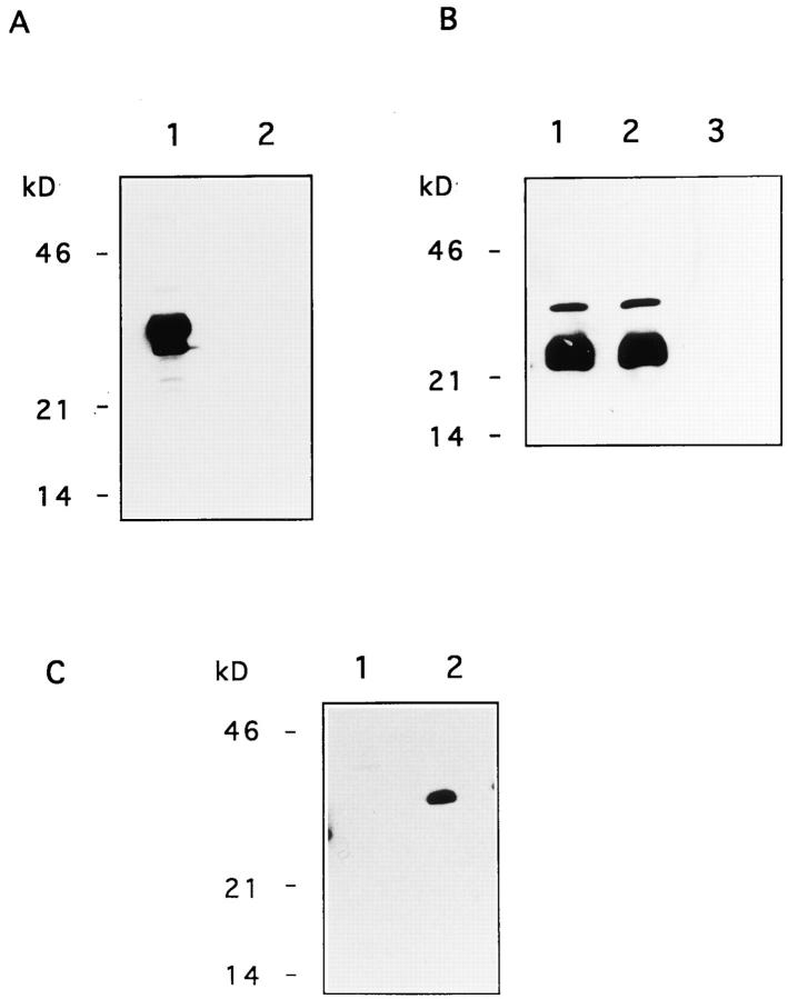
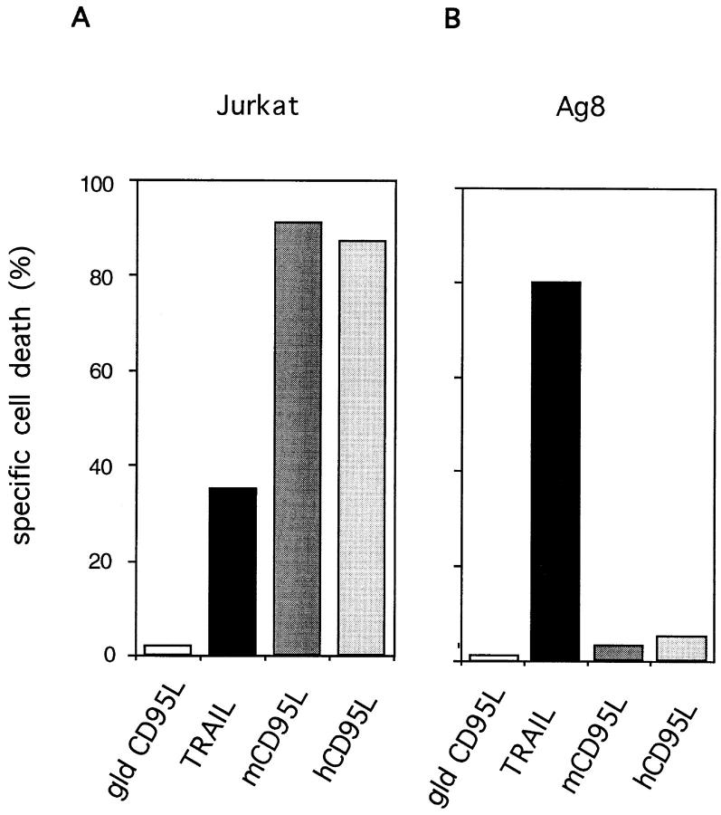
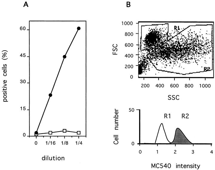
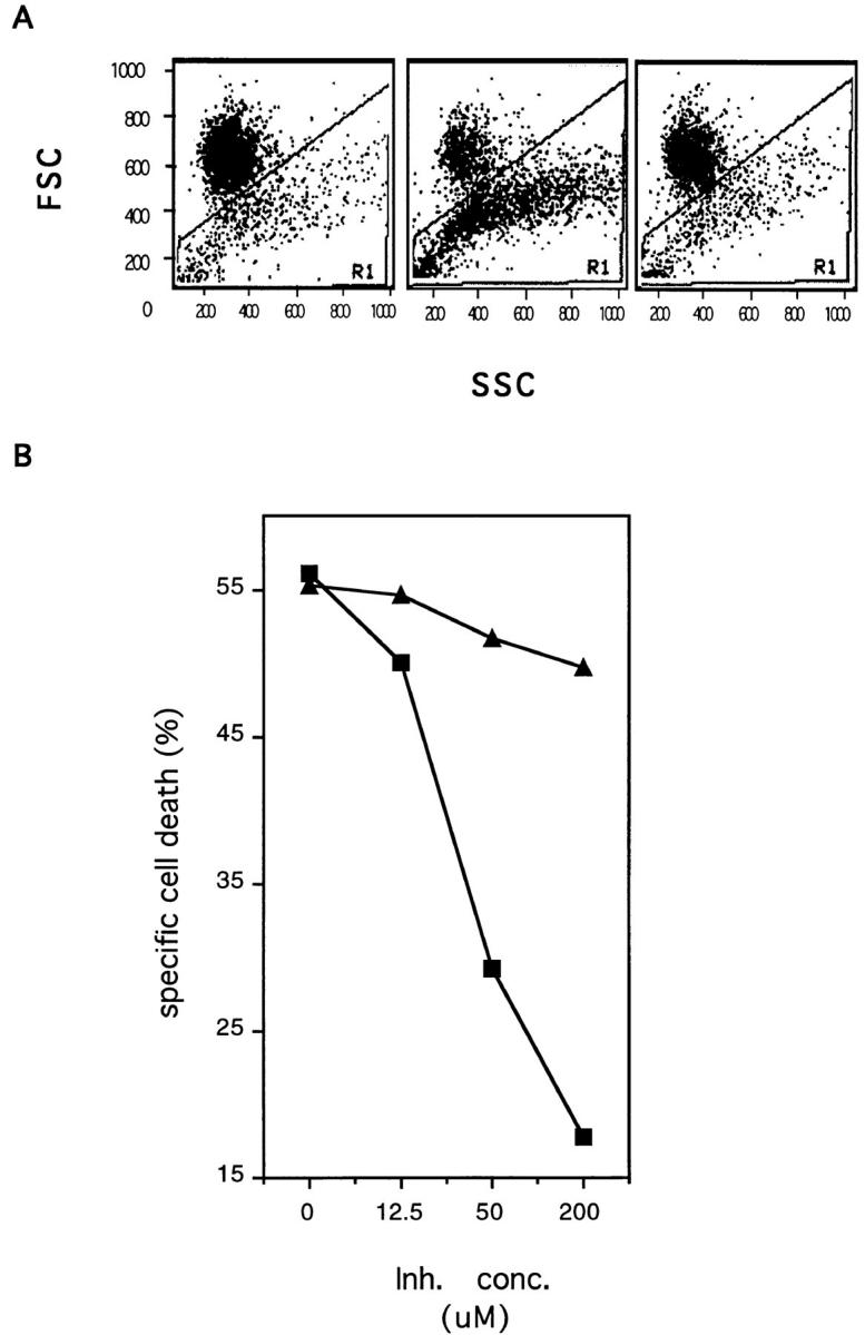
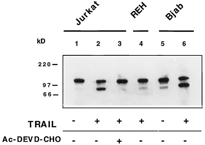
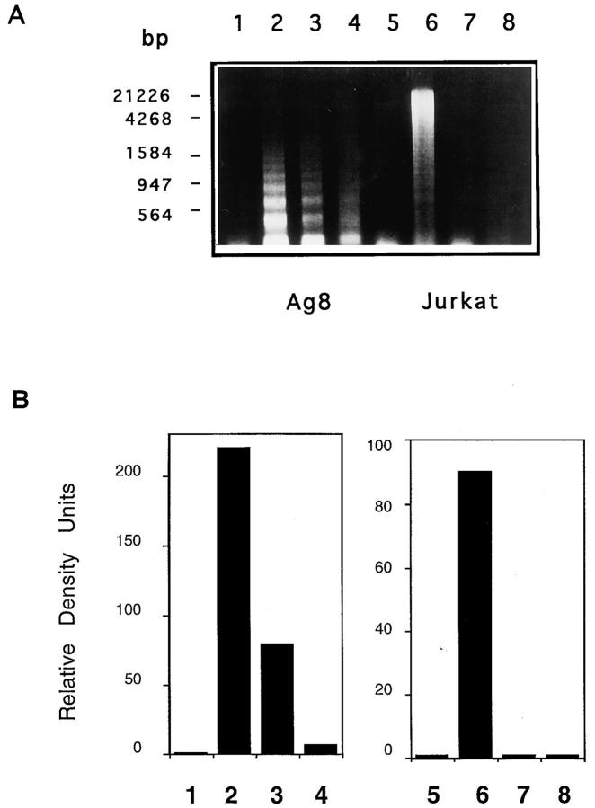
The Fas/APO-1/CD95 ligand (CD95L) and the recently cloned TRAIL ligand belong to the TNF-family and share the ability to induce apoptosis in sensitive target cells. Little information is available on the degree of functional redundancy between these two ligands in terms of target selectivity and intracellular signalling pathway(s). To address these issues, we have expressed and characterized recombinant mouse TRAIL. Specific detection with newly developed rabbit anti-TRAIL antibodies showed that the functional TRAIL molecule released into the supernatant of recombinant baculovirus-infected Sf9 cells is very similar to that associated with the membrane fraction of Sf9 cells. CD95L resistant myeloma cells were found to be sensitive to TRAIL, displaying apoptotic features similar to those of the CD95L- and TRAIL-sensitive T leukemia cells Jurkat. To assess if IL-1beta-converting enzyme (ICE) and/or ICE-related proteases (IRPs) (caspases) are involved in TRAIL-induced apoptosis of both cell types, peptide inhibition experiments were performed. The irreversible IRP/caspase-inhibitor Ac-YVAD-cmk and the reversible IRP/caspase-inhibitor Ac-DEVD-CHO blocked the morphological changes, disorganization of plasma membrane phospholipids, DNA fragmentation, and loss of cell viability associated with TRAIL-induced apoptosis. In addition, cells undergoing TRAIL-mediated apoptosis displayed cleavage of poly(ADP)-ribose polymerase (PARP) that was completely blocked by Ac-DEVD-CHO. These results indicate that TRAIL seems to complement the activity of the CD95 system as it allows cells, otherwise resistant, to undergo apoptosis triggered by specific extracellular ligands. Conversely, however, induction of apoptosis in sensitive cells by TRAIL involves IRPs/caspases in a fashion similar to CD95L. Thus, differential sensitivity to CD95L and TRAIL seems to map to the proximal signaling events associated with receptor triggering.


Figures

Figure 1
Figure 1
Immunoblot analysis of recombinant mouse TRAIL. (A) Whole cell lysates of Sf9 cells expressing recombinant mouse TRAIL (lane 1) or mouse CD95L (lane 2) were tested with purified rabbit anti-TRAIL Ab. Molecular weight markers are indicated. HRPO-labeled mouse anti–rabbit IgG antibodies were used as the detecting reagent. No reactivity was present with purified rabbit IgG. (B) Whole cell lysates of Sf9 cells expressing recombinant mouse TRAIL (lane 1) or mouse CD95L (lane 2) and cell lysates of non-infected Sf9 cells (lane 3) were tested with the mouse mAb SM22 to a baculovirus-related product. Molecular weight markers are indicated. HRPO-labeled rabbit anti–mouse IgG antibodies were used as the detecting reagent. No reactivity was present with an isotype-matched control mAb. (C) SN of Sf9 cells expressing mouse TRAIL (lane 2) or mouse CD95L (lane 1) were tested with purified rabbit anti-TRAIL antibody. Molecular weight markers are indicated. HRPO-labeled mouse anti–rabbit IgG antibodies were used as the detecting reagent. No reactivity was found with purified rabbit IgG.
Figure 2
Figure 2
Target cells resistant to mouse CD95L are sensitive to mouse TRAIL. CD95L-sensitive human leukemia Jurkat cells (Jurkat) (A) and CD95L-resistant mouse myeloma cells Ag8 (Ag8) (B) were incubated overnight with SN from Sf9 cells expressing mouse gld CD95L (gld CD95L), mouse TRAIL (TRAIL), mouse wild-type CD95L (mCD95L), and human CD95L (hCD95L). Results are expressed as percent specific cell death. Background apoptosis of the target cells incubated with SN of mock-infected Sf9 cells was <15%.
Figure 3
Figure 3
Induction of apoptosis and DNA fragmentation in Ag8 cells by recombinant mouse TRAIL. (A) The human T leukemia cells Jurkat (Jurkat) (left panels), the mouse myeloma cells Ag8 (Ag8) (central panels), and the mouse T hybridoma cells 2H11 (TcHy) (right panels) were incubated with SN from Sf9 cells expressing mouse TRAIL (TRAIL) (upper panels), or mouse wildtype CD95L (CD95L) (lower panels). Results are presented as forward/side scatter analysis of cellular morphology. Apoptotic cells are shown in gate R1. (B) Soluble DNA was extracted from Ag8 (Ag8) (lanes 1–3) and Jurkat cells (Jurkat) (lanes 4–6) (0.5 × 10⁶ cells/lane) incubated with SN from mock-infected Sf9 (lanes 1 and 4) or from Sf9 cells expressing mouse CD95L (lanes 2 and 5) or mouse TRAIL (lanes 3 and 6). Extracted DNA was separated on an agarose gel and visualized by ethidium bromide staining. Molecular mass markers (MM) are shown. No soluble DNA was extracted from TRAIL-resistant T hybridoma cells incubated with either ligand.
Figure 3
Figure 3
Induction of apoptosis and DNA fragmentation in Ag8 cells by recombinant mouse TRAIL. (A) The human T leukemia cells Jurkat (Jurkat) (left panels), the mouse myeloma cells Ag8 (Ag8) (central panels), and the mouse T hybridoma cells 2H11 (TcHy) (right panels) were incubated with SN from Sf9 cells expressing mouse TRAIL (TRAIL) (upper panels), or mouse wildtype CD95L (CD95L) (lower panels). Results are presented as forward/side scatter analysis of cellular morphology. Apoptotic cells are shown in gate R1. (B) Soluble DNA was extracted from Ag8 (Ag8) (lanes 1–3) and Jurkat cells (Jurkat) (lanes 4–6) (0.5 × 10⁶ cells/lane) incubated with SN from mock-infected Sf9 (lanes 1 and 4) or from Sf9 cells expressing mouse CD95L (lanes 2 and 5) or mouse TRAIL (lanes 3 and 6). Extracted DNA was separated on an agarose gel and visualized by ethidium bromide staining. Molecular mass markers (MM) are shown. No soluble DNA was extracted from TRAIL-resistant T hybridoma cells incubated with either ligand.
Figure 4
Figure 4
Induction of plasma membrane phospholipid disorganization by recombinant mouse TRAIL. The T leukemia cells Jurkat were incubated overnight with serial dilutions of SN from Sf9 cells expressing mouse TRAIL (filled circles) or mouse gld CD95L (empty squares). Results are expressed as percent cells with high uptake of the lipophilic dye MC540 (A). In B, results are presented as forward/side scatter analysis of cells incubated with recombinant TRAIL. MC540 uptake is shown for cells with nonapoptotic morphology (gate R1) (white histogram) and for cells with apoptotic morphology (gate R2) (gray histogram). No difference in cell morphology was observed in Jurkat cells exposed to TRAIL in the presence or absence of MC540.
Figure 5
Figure 5
The IRP/caspase-inhibitor Ac-YVAD-cmk blocks TRAILinduced apoptosis in mouse myeloma cells. (A) The mouse myeloma cells Ag8 were incubated overnight with SN of Sf9 cells expressing mouse TRAIL (central and right panel) in the presence of the peptide inhibitor Ac-YVAD-cmk (200 μM) (right panel) or an equal concentration of vehicle (dmso) (central panel). As a specificity control Ag8 cells were incubated with SN of mockinfected Sf9 cells (left panel). Results are presented as forward/ side scatter analysis of cellular morphology. Apoptotic cells are shown in gate R1. (B) Ag8 cells were incubated overnight with SN of Sf9 cells expressing mouse TRAIL in the presence of increasing concentrations of the peptide inhibitor Ac-YVAD-cmk (squares). Equal concentrations of vehicle (triangles) were used as a specificity control. Results are expressed as percent specific cell death. Background apoptosis of target cells was <15%.
Figure 6
Figure 6
The IRP/caspase-inhibitor Ac-YVAD-cmk blocks TRAILinduced apoptosis in human leukemia cells. (A) The human leukemia cells Jurkat were incubated overnight with SN of Sf9 cells expressing mouse TRAIL in the presence of increasing concentrations of the peptide inhibitor Ac-YVAD-cmk (circles). Equal concentrations of vehicle (squares) were used as a specificity control. Results are expressed as percent specific cell death. Background apoptosis of target cells was less than 15%. (B) Jurkat cells were incubated for 8 h with SN of Sf9 cells expressing mouse TRAIL in the presence of increasing concentrations of the peptide inhibitor Ac-YVAD-cmk (circles). Equal concentrations of vehicle (squares) were used as a specificity control. Results are expressed as percent of cells with high uptake of the MC540 dye.
Figure 7
Figure 7
The IRP/caspase inhibitor Ac-DEVD-CHO blocks TRAIL-induced PARP cleavage. Whole cell lysates of Jurkat cells incubated with SN of Sf9 cells expressing mouse TRAIL (lanes 2–3) or of mock-infected Sf9 cells (lane 1) in the presence (lane 3) or absence of Ac-DEVD-CHO (lanes 1–2) were tested with the anti-PARP Ab. The TRAIL-resistant REH (lane 4) and the TRAIL-sensitive BJAB cells (lanes 5–6) incubated with (lanes 4 and 6) or without TRAIL (lane 5) were used as specificity controls. Molecular markers are indicated. HRPO-labeled goat anti–mouse IgG Ab were used as the detecting reagent.
Figure 8
Figure 8
The Ac-YVAD-cmk and Ac-DEVD-CHO peptide inhibitors block TRAIL-induced DNA fragmentation in mouse and human cells. (A) Soluble DNA was extracted from mouse myeloma cells Ag8 (lanes 1–4) (Ag8) or from the human leukemia cells Jurkat (lanes 5–8) (Jurkat) (0.5 × 10⁶ cells/lane) incubated with SN from mock-infected Sf9 (lanes 1 and 5) or with SN from TRAIL-expressing Sf9 cells (lanes 2–4 and 6–8) in the presence of the peptide inhibitor Ac-YVAD-cmk (lanes 3 and 7) or Ac-DEVD-CHO (lanes 4 and 8) (200 μM). An equal concentration of vehicle (dmso) was used as a specificity control (lanes 2 and 6). Extracted DNA was separated on an agarose gel and visualized by ethidium bromide staining. Molecular mass markers (MM) are shown. In B, results are shown as relative density of fragmented DNA in each lane (relative density units) as determined by densitometric analysis.

References

    1. Alnemri ES, Livingston DJ, Nicholson DW, Salvesen G, Thornberry NA, Wong WW, Yuan J. Human ICE/CED-3 protease nomenclature. Cell. 1996;87:171. - PubMed
    1. Ashkenaz J, Werb Z. Proteolysis and the biochemistry of life-ordeath decisions. J Exp Med. 1996;183:1947–1951. - PMC - PubMed
    1. Boldin MP, Varfolomeev EE, Pancer Z, Mett IL, Camonis JH, Wallach D. A novel protein that interacts with the death domain of FAS/ APO-1 contains a sequence motif related to the death domain. J Biol Chem. 1995;270:7795–7798. - PubMed
    1. Boldin MP, Goncharov TM, Goltsev YV, Wallach D. Involvement of MACH, a novel MORT1/FADD-interacting protease, in Fas/APO-1- and TNF receptor-induced cell death. Cell. 1996;85:803–815. - PubMed
    1. Brunner T, Mogil RJ, LaFace D, Yoo NJ, Mahboubi A, Echeverry F, Martin SJ, Force WR, Lynch DH, Ware CF, et al. Cell-autonomous Fas (CD95)/Fas-ligand interaction mediates activation-induced apoptosis in T-cell hybridoma. Nature (Lond) 1995;373:441–444. - PubMed

Publication types

MeSH terms