. 1997 Apr 15;94(8):3542-7.
doi: 10.1073/pnas.94.8.3542.

Nuclear antisense RNA induces extensive adenosine modifications and nuclear retention of target transcripts

Affiliations

Nuclear antisense RNA induces extensive adenosine modifications and nuclear retention of target transcripts

M Kumar et al. Proc Natl Acad Sci U S A. .

Abstract

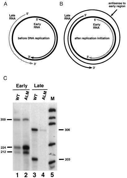
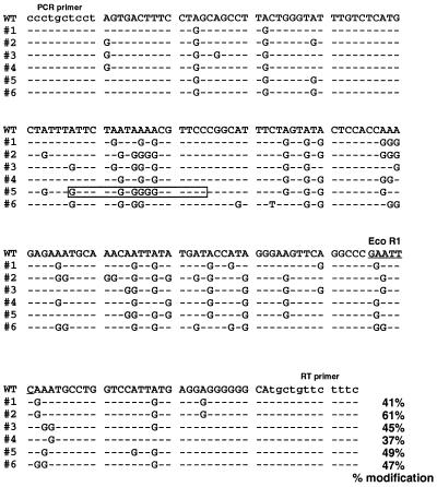
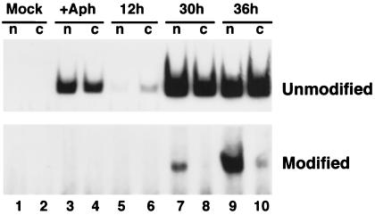
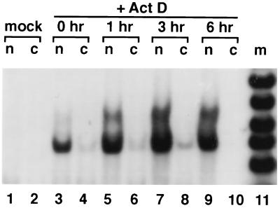
Antisense RNA may regulate the expression of a number of eukaryotic genes, but little is known about its prevalence or mechanism of action. We have used a model system in which antisense control can be studied both genetically and biochemically. Late in polyoma virus infection, early-strand mRNA levels are down-regulated by nuclear antisense RNA from the late strand. Analysis of early-strand transcripts isolated late in infection revealed extensive base modifications. In many transcripts almost half of the adenosines were altered to inosines or guanosines. These results suggest modification of RNA duplexes by double-stranded RNA adenosine deaminase or a related enzyme. Probes that detect only modified RNAs revealed that these molecules are not highly unstable, but accumulate within the nucleus and are thus inert for gene expression. Antisense-induced modifications can account for most or all of the observed regulation, with the lowered levels of early-strand RNAs commonly observed late in infection resulting from the fact that many transcripts are invisible to standard hybridization probes. This work suggests that similar antisense-mediated control mechanisms may also operate under physiological conditions in uninfected eukaryotic cells, and leads to the proposal that there is a novel pool of nuclear RNAs that cannot be seen with many molecular probes heretofore used.


Figures

Figure 1
Figure 1
Temporal regulation of polyoma virus transcript levels. (A) During the early phase of viral infection, early-strand transcripts accumulate preferentially over late-strand transcripts. Late-strand transcripts are processed inefficiently and are relatively unstable. Prior to DNA replication the ratio of late-strand to early-strand RNAs is less than 1:10. (B) During the late phase of infection, after the onset of DNA replication, late-strand transcripts are more abundant than early-strand transcripts. Transcription termination is inefficient during this period, allowing RNA polymerase II to encircle the genome multiple times. The resulting multigenomic transcripts contain sequences complementary to early-strand transcripts and thus have the potential to act as natural antisense regulators within the nucleus. Hatched lines denote unstable transcripts. (C) The antisense effect. Total cell RNA was harvested 48 hr after transfection with either wild-type genomes (WT) or mutant ALM (which contains a 51-bp deletion in the viral late region, which leads to unstable late-strand primary transcripts) genomes, and analyzed for early-strand (lanes 1 and 2) and late-strand (lanes 3 and 4) transcripts using specific riboprobes that span splicing junctions. Positions of expected early-strand and late-strand bands from wild-type and marked constructs are indicated. M, molecular weight markers produced by digesting pUC18 with MspI. Quantitation using a Packard InstantImager revealed that mutant ALM produced less than 10% as much late-strand RNA as wild type, and overexpressed early-strand RNAs by a factor of 5.
Figure 2
Figure 2
Analysis of RT-PCR products. A region of the early sense strand was reverse-transcribed and amplified by RT-PCR using primers that would hybridize to the RNA irrespective of adenosine modifications. The primer used for reverse transcription was 5′-AGAAAGAACAGCA-3′. The second primer used for PCR amplification was 5′-TCCCCCTGCTCCT-3′. The amplified product was gel-purified and subjected to EcoRI digestion. The bands were separated by agarose gel electrophoresis. Lane 1, marker. Lane 2, control experiment where cells were treated with aphidicolin to block DNA replication. RNase protection analysis revealed the presence of sufficient amounts of early-strand RNAs, while late-strand RNAs were barely detectable (data not shown). These cells express very low levels of late-strand transcripts. Lanes 3–5, results of three independent experiments. Bands corresponding to the EcoRI-digested and resistant fragments are indicated. As seen in the figure, a considerable amount of RT-PCR amplified product was resistant to EcoRI as compared with the control. The EcoRI-resistant band was purified, cloned into pBluescript SK(+), and sequenced.
Figure 3
Figure 3
Sequence comparison of six modified clones with that of genomic DNA. WT, sequence of the wild-type polyoma genome. #1–#6, sequences of the modified clones. Only the modified bases are shown in the six clones; other sequences were identical, except as noted. Lowercase letters indicate primer binding sites. The EcoR1 site is underlined, and the percentage of the modified adenosines is indicated for each sequence. A synthetic DNA oligodeoxynucleotide complementary to the sequence within the boxed region was used for the experiments described in Fig. 4.
Figure 4
Figure 4
Determination of the intracellular fate of modified RNAs. Mouse NIH 3T3 cells were mock-infected, or infected with wild-type polyoma virus for the times indicated. At the indicated times, nuclear and cytoplasmic RNAs were isolated, and 5 μg of each fraction used for RT-PCR amplification, as in Fig. 2. Reverse transcription was performed using a primer containing the sequence complementary to the modified or unmodified sequence shown in the boxed area in Fig. 3 (5′-GGAACGCCCCACTAGAAC-3′). Subsequent PCR amplification used, in addition, the “PCR primer” noted in lowercase letters in Fig. 3. The PCR products corresponding to amplification of modified or unmodified RNAs are 74 bp in length, and are indicated in the figure.
Figure 5
Figure 5
Actinomycin D time course measurements of RNA stabilities. Nuclear and cytoplasmic RNAs were isolated at various times after treatment of cells with actinomycin D, and 5 μg of each fraction was used for RT-PCR amplification, as in Fig. 2, using the same primers as in Fig. 4.

References

    1. Inouye M. Gene. 1988;72:25–34. - PubMed
    1. Simons R W. Gene. 1988;72:35–44. - PubMed
    1. Tosic M, Roach A, de Rivaz J-C, Dolivo M, Matthieu J-M. EMBO J. 1990;9:401–406. - PMC - PubMed
    1. Volk R, Köster M, Pöting A, Hartmann L, Knöchel W. EMBO J. 1989;8:2983–2988. - PMC - PubMed
    1. Williams T, Fried M. Nature (London) 1986;322:275–279. - PubMed