. 1997 Apr 15;94(8):3571-5.
doi: 10.1073/pnas.94.8.3571.

A structural model for GroEL-polypeptide recognition

Affiliations

A structural model for GroEL-polypeptide recognition

A M Buckle et al. Proc Natl Acad Sci U S A. .

Abstract

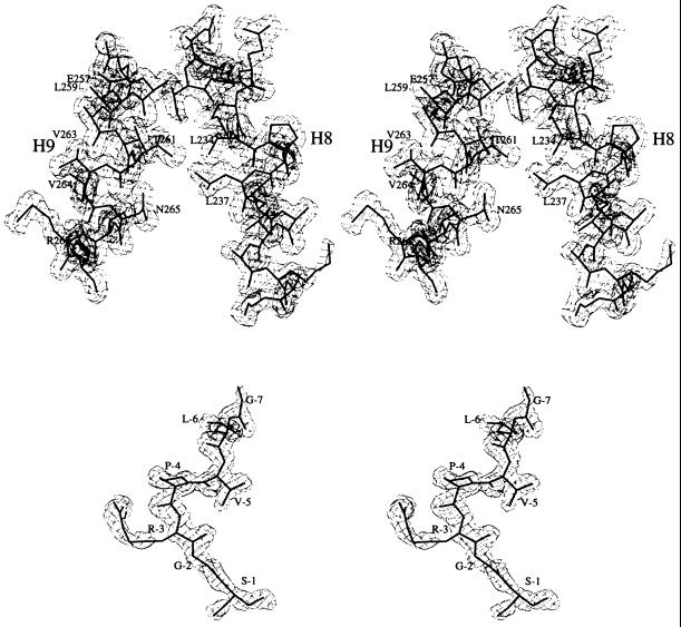
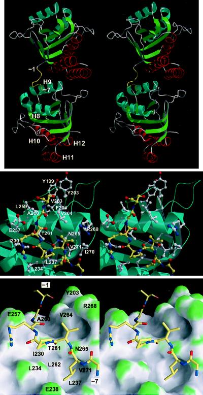
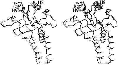
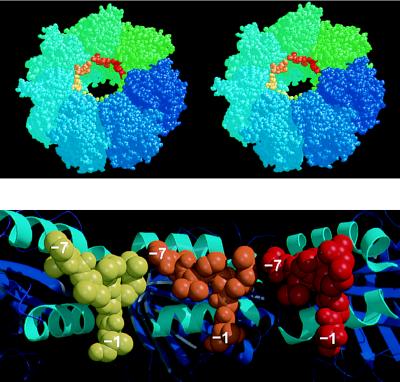
A monomeric peptide fragment of GroEL, consisting of residues 191-376, is a mini-chaperone with a functional chaperoning activity. We have solved the crystal structure at 1.7 A resolution of GroEL(191-376) with a 17-residue N-terminal tag. The N-terminal tag of one molecule binds in the active site of a neighboring molecule in the crystal. This appears to mimic the binding of a peptide substrate molecule. Seven substrate residues are bound in a relatively extended conformation. Interactions between the substrate and the active site are predominantly hydrophobic, but there are also four hydrogen bonds between the main chain of the substrate and side chains of the active site. Although the preferred conformation of a bound substrate is essentially extended, the flexibility of the active site may allow it to accommodate the binding of exposed hydrophobic surfaces in general, such as molten globule-type structures. GroEL can therefore help unfold proteins by binding to a hydrophobic region and exert a binding pressure toward the fully unfolded state, thus acting as an "unfoldase." The structure of the mini-chaperone is very similar to that of residues 191-376 in intact GroEL, so we can build it into GroEL and reconstruct how a peptide can bind to the tetradecamer. A ring of connected binding sites is noted that can explain many aspects of substrate binding and activity.


Figures

Figure 1
Figure 1
2Fo-Fc electron density map calculated with SIGMAA coefficients (23), contoured at 1σ (σ is the root-mean-square deviation from the mean electron density in the unit cell). Superimposed is the refined atomic model. (Upper) Helices H8 and H9. (Lower) The N-terminal tag. Drawn with the bobscript (extensions to the program molscript; ref. 24).
Figure 2
Figure 2
(Top) Stereo cartoon representation of the structure of the mini-chaperone (GroEL191–376), showing the interaction between the N-terminal tag and a neighboring molecule in the crystal lattice (related by a crystallographic two-fold screw operation along the c axis, positioned approximately vertical and in the plane of the paper). The N-terminal tag (residues −1 to −7) is colored yellow. (Middle) Close-up of peptide-binding site interactions, in stereo. The peptide is represented by yellow bonds; neighboring residues are represented by white bonds. Hydrogen bonds are represented by broken white lines. Drawn with bobscript (extensions to the program molscript; ref. 24) and raster3d (25). (Lower) As in Middle but showing the molecular surface of the mini-chaperone. The surface is colored according to surface curvature to highlight concave surface pockets. Convex, concave, and flat surfaces are colored green, grey, and white, respectively. Residues underlying the surface are labeled. Drawn with grasp (26). All three figures show the model in approximately the same orientation.
Figure 3
Figure 3
Stereo representation of the overlay of Cα atoms of the apical domain in intact GroEL (residues 191–376; broken bonds), mini-chaperone GroEL191–376 (thick bonds), and mini-chaperone GroEL191–345 (thin bonds) (17). Structures were fitted using backbone atoms from β-sheet residues. Drawn with molscript (24).
Figure 4
Figure 4
(Upper) Stereoview of one heptameric ring of the GroEL tetradecamer showing the position of the N-terminal tag bound to each apical domain, near the opening to the central cavity. This model is generated by the superposition of the mini-chaperone GroEL(191–376) with each corresponding apical domain (residues 191–376) in intact GroEL (the second ring of the GroEL cylinder, generated by a two-fold symmetry operation, is not shown, but stacks against the underside of the drawn ring). GroEL subunits are colored around the ring going from blue to green. Superimposed “bound peptides” are colored from green to red. Drawn with rasmol (32). (Lower) Cross-section of the model shown in Upper looking directly at the inner wall of the cavity, and showing the apical domains (cartoon with helices H8 and H9 colored cyan) from three subunits with modeled peptide (shown as space-filling models colored yellow, orange, and red, respectively). Drawn with molscript (24) and raster3d (25). Each of the separate peptides (residues −1 to −7) could be linked together by small fragments of peptides so that a longer peptide could bind from one contiguous site to the next.

References

    1. Braig K, Otwinowski Z, Hegde R, Boisvert D C, Joachimiak A, Horwich A L, Sigler P B. Nature (London) 1994;371:578–86. - PubMed
    1. Braig K, Adams P D, Brünger A T. Nat Struct Biol. 1995;2:1083–1094. - PubMed
    1. Landry S J, Gierasch L M. Biochemistry. 1991;30:7359–7362. - PubMed
    1. Landry S J, Jordan R, McMacken R, Gierasch L M. Nature (London) 1992;355:455–457. - PubMed
    1. Hayer-Hartl M K, Ewbank J J, Creighton T E, Hartl F-U. EMBO J. 1994;13:3192–3202. - PMC - PubMed