. 1997 Apr 15;94(8):3584-9.
doi: 10.1073/pnas.94.8.3584.

Crystal structure of Lyme disease antigen outer surface protein A complexed with an Fab

Affiliations

Crystal structure of Lyme disease antigen outer surface protein A complexed with an Fab

H Li et al. Proc Natl Acad Sci U S A. .

Abstract

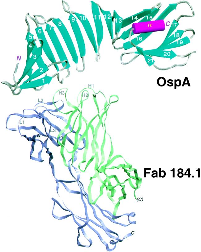
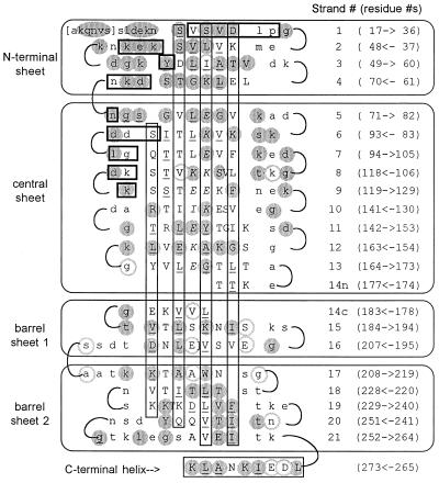
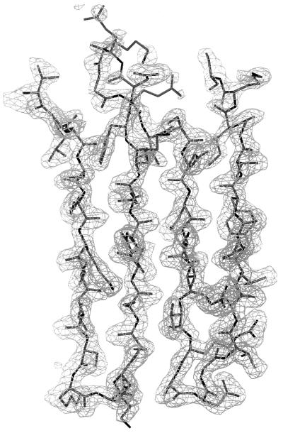
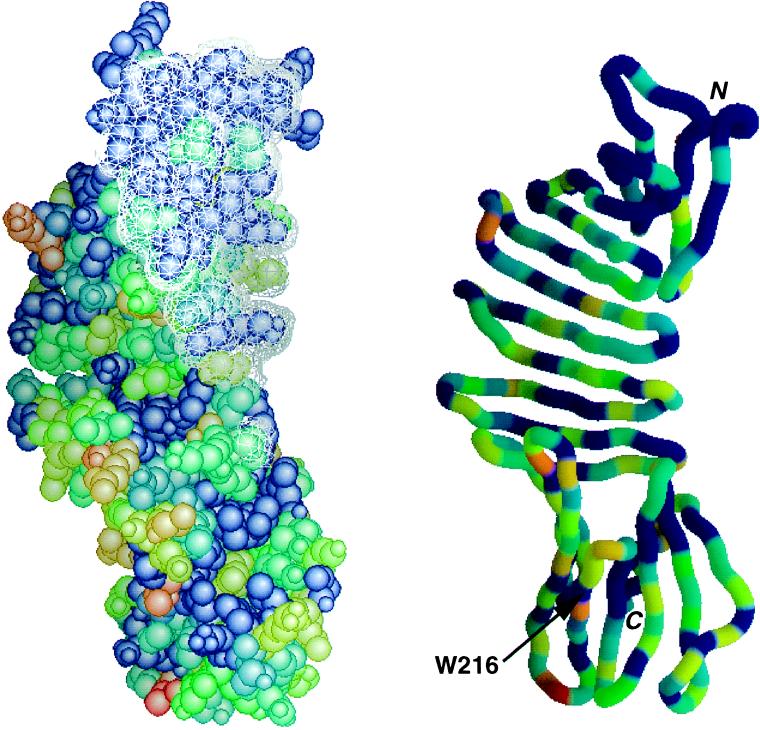
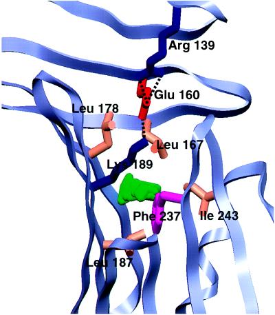
OspA (outer surface protein A) is an abundant immunogenic lipoprotein of the Lyme disease spirochete Borrelia burgdorferi. The crystal structure of a soluble recombinant form of OspA was solved in a complex with the Fab fragment of mouse monoclonal antibody 184.1 and refined to a resolution of 1.9 A. OspA has a repetitive antiparallel beta topology with an unusual nonglobular region of "freestanding" sheet connecting globular N- and C-terminal domains. Arrays of residues with alternating charges are a predominant feature of the folding pattern in the nonglobular region. The 184.1 epitope overlaps with a well conserved surface in the N-terminal domain, and a hydrophobic cavity buried in a positively charged cleft in the C-terminal domain is a potential binding site for an unknown ligand. An exposed variable region on the C-terminal domain of OspA is predicted to be an important factor in the worldwide effectiveness of OspA-based vaccines.


Figures

Figure 1
Figure 1
Schematic view of the OspA–Fab 184.1 complex. OspA is displayed with its secondary structural elements, β-strands (cyan, numbered 1–21) and α-helix (magenta, labeled α), connected by grey turns or loops. The front face of the central sheet, as defined in the text, is seen in this view. Fab 184.1 is represented by violet and green ribbons for the light and heavy chains, respectively. The three complementarity determining region (CDR) loops on each chain are displayed as thin ropes and labeled (e.g., L1 for CDR1 of the light chain). The figure was generated with setor (21).
Figure 2
Figure 2
Two-dimensional representation of the OspA folding pattern. The view is of the front face of the central β-sheet, with other β-sheets “uncurled” to form a single pseudocontinuous sheet. Residues in secondary structural elements (β-strands or α-helix) are represented by uppercase type; residues in turns are in lowercase type. Residues in brackets at the N terminus are predicted to be present in recombinant OspA but are not seen in the crystal. The four β-sheets formed by the 21 antiparallel β-strands are indicated by labeled rectangles enclosing sections of sequence, and the C-terminal α-helix is indicated at the bottom. For each line, the β-strand number is indicated in parentheses to the right, followed by the numerical range and direction of displayed residues. Vertical rectangles indicate residues with side chains extending behind the plane of the figure, residues with uppercase type outside the rectangles extend toward the viewer. Residues with exposed surface areas less than 10 Å2 are considered buried and are underlined. In general, buried residues are front-facing between strands 1 and 9 and rear-facing between strands 11 to 21, corresponding to the folding of N- and C-terminal domains on opposite sides of the central sheet. Strand 14 has main-chain hydrogen bonds to both the central sheet (14n) and to barrel sheet 1 (14c). Residues that contact Fab 184.1 are horizontally boxed (heavy lines); residues in the charge arrays are indicated in italic type. Residues strictly conserved in 49 OspA sequences (see Fig. 4) are within shaded circles; highly variable residues are within circles with shaded outlines.
Figure 3
Figure 3
Electron density for antiparallel β-strands 8–11 in the central sheet of OspA. The (2FobsFcalc), model phase map is contoured at 1.0 σ (mesh). The thick-wire model extends from Thr-108 at the upper left through two β-hairpins to Ser-152 at the upper right. Note poor density for the flexible loop (upper center) between β-strands 9 and 10, typical for the larger more-flexible loops on the upper edge of the central sheet.
Figure 4
Figure 4
Conservation map for OspA, with epitope of Fab 184.1 superimposed. Forty-nine OspA sequences available in GenBank in February 1996 were aligned, and the results were encoded on a 0–10 conservation scale using alscript (38). OspA was then color-coded according to conservation, with red representing the most variable residues and blue representing strictly conserved residues. Similar results are obtained when the 19 available OspB sequences are also included (data not shown). (Left) Space-filling model of OspA, with the surface made inaccessible by Fab 184.1 indicated as a white mesh. (Right) Ribbon model in same orientation, with same coloring scheme. Included were sequences from the genospecies B. burgdorferi sensu stricto, Borrelia garinii, Borrelia afzelii, Borrelia japonica, and Borrelia andersonii (16, 17). The figure was made with grasp (39).
Figure 5
Figure 5
Close-up of the C-terminal domain potential ligand binding pocket. The main chain of OspA is shown as a light-blue ribbon; residues forming the charge trio (Arg-139, Glu-160, and Lys-189) or lining the 34-Å3 cavity (Leu-167, Leu-178, Leu-187, Phe-237, and Ile-243) are shown in colors representative of side-chain type. The cavity, represented in green, was calculated by using the University of Groningen biomol suite. The figure was generated with setor (21).

References

    1. Barbour A G, Fish D. Science. 1993;260:1610–1616. - PubMed
    1. Barbour A G, Tessier S L, Todd W J. Infect Immun. 1983;41:795–804. - PMC - PubMed
    1. Howe T R, Mayer L W, Barbour A G. Science. 1985;227:645–646. - PubMed
    1. Howe T R, LaQuier F R, Barbour A G. Infect Immun. 1986;54:207–212. - PMC - PubMed
    1. Brusca J S, McDowall A W, Norgard M V, Radolf J D. J Bacteriol. 1991;173:8004–8008. - PMC - PubMed

Publication types

MeSH terms

Associated data