. 1997 Apr 15;94(8):3627-32.
doi: 10.1073/pnas.94.8.3627.

Transduction of interleukin-2 antiapoptotic and proliferative signals via Akt protein kinase

Affiliations

Transduction of interleukin-2 antiapoptotic and proliferative signals via Akt protein kinase

N N Ahmed et al. Proc Natl Acad Sci U S A. .

Abstract

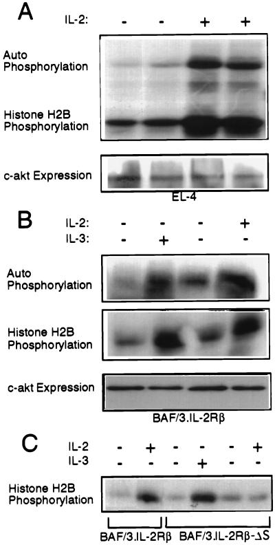
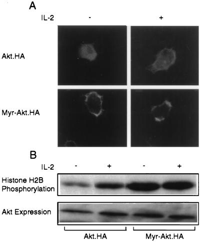
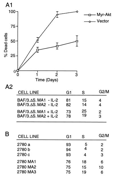
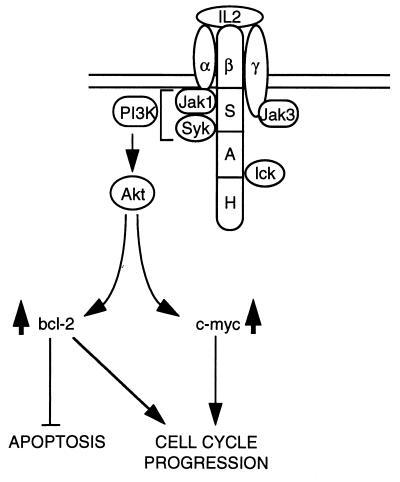
The interleukin-2 (IL-2) receptor (IL-2R) is composed of three subunits. Of these, IL-2Ra is required for high-affinity IL-2 binding, while IL-2R beta and IL-2R gamma(c) are required for the transduction of IL-2-generated signals. Signals transduced via the S region of the IL-2R beta (amino acids 267-322) in BAF/3 cells activate the phosphatidylinositol 3-kinase (PI3-kinase) and induce the expression of Bcl-2 and c-myc. Through the induction of Bcl-2, IL-2 inhibits apoptosis and through the combination of Bcl-2 and c-myc it stimulates progression through the cell cycle. Here we show that the protein kinase encoded by the Akt proto-oncogene is activated by IL-2. Akt activation by IL-2 depends on PI3-kinase signals transduced via the S region of the IL-2R beta and is linked to the translocation of Akt to the cell membrane. Expression of catalytically active Akt mutants in BAF/3 cells expressing IL-2R beta[A0]delta S promotes the expression of Bcl-2 and c-myc, inhibits apoptosis induced by IL-3 deprivation or staurosporine, and stimulates cell cycle progression. The same mutants also stimulate cell cycle progression in 2780a, an IL-2-dependent T cell line that undergoes G1 arrest rather than apoptosis after IL-2 deprivation. The activation of Akt by IL-2 via the PI3-kinase and the rescue of the PI3-kinase-mediated antiapoptotic and proliferative IL-2 signals by catalytically active Akt indicate that these signals are transduced by Akt.


Figures

Figure 1
Figure 1
Akt is activated by IL-2 and IL-3 in EL4·IL-2 cells and BAF/3 cells expressing IL-2Rβ, but not IL-2Rβ·ΔS. (A) Five × 106 EL4·IL-2 (ATCC, TIB 181) cells, cultured at a concentration of 0.5 × 10⁶ cells per ml, were serum-starved for 16 h. Some of the starved cultures then were stimulated with IL-2 (100 units/ml) for 10 min as indicated. (Upper) In vitro kinase assays of Akt immunoprecipitated from Nonidet P-40 lysates of unstimulated or IL-2-stimulated EL4 cells, using the anti-Akt-CT antibody. (Lower) Anti-Akt-CT Western blot of the immunoprecipitates used in the kinase assay above. (B) Five × 106 BAF/3 cells stably transfected with the expression construct pdKCR-IL-2Rβ (3) cultured at a concentration of 0.5 × 10⁶ cells per ml, were starved of IL-3 for 16 h. (Top and Middle) In vitro kinase assays carried out on anti-Akt immunoprecipitates of unstimulated and IL-2 (100 units/ml) or IL-3 (50 units/ml) stimulated cell lysates. (Bottom) Expression of c-akt as determined by probing Western blots of total cell lysates with the anti-Akt-CT antibody. (C) BAF/3 cells (5 × 10⁶) stably expressing the IL-2Rβ or the IL-2Rβ·ΔS mutant, cultured at the concentration of 0.5 × 10⁶ cells per ml, were transiently transfected with HA·Akt. Twenty-four hours after transfection, the cells were starved of IL-2 and IL-3 for 16 h. Subsequently, they were stimulated with IL-2 or IL-3 for 10 min as indicated. In vitro kinase assays were carried out on HA[A0]Akt immunoprecipitated from Nonidet P-40 cell lysates.
Figure 2
Figure 2
Akt activation by IL-2 is linked to the translocation of Akt to the cell membrane. (A) 107 EL4·IL-2 cells transiently transfected with carboxyl-terminally tagged Akt (Akt·HA) or myristylated Akt (Myr-Akt·HA) expression constructs and stained with the anti-HA monoclonal antibody 12CA5 and an anti-mouse fluorescein isthiocyanate-conjugated secondary antibody before and after treatment with IL-2. (Upper) Cells transfected with wild-type Akt. (Lower) Cells transfected with myristylated Akt. (B) In vitro kinase assays of Akt·HA and myristylated Akt·HA immunoprecipitated from Nonidet P-40 lysates of the cells described in A.
Figure 3
Figure 3
Myristylated Akt inhibits apoptosis and cell cycle arrest induced by growth factor withdrawal in growth factor dependent lymphoid cells. (A1) Expression of myristylated Akt inhibits apoptosis induced by IL-2/IL-3 withdrawal in IL-2Rβ·ΔS expressing BAF/3 cells. Three independent cultures of BAF/3-IL-2Rβ·ΔS cells, stably transfected with a CMV-6 construct of myristylated Akt or the CMV-6 vector alone, were cultured in IL-2 and IL-3 deficient media at the concentration of 0.5 × 10⁶ cells per ml. The percentage of dead cells over time was calculated from the number of live and dead cells counted daily as described in the text. (A2) Myristylated Akt inhibits G1 arrest of BAF/3-IL-2Rβ·ΔS cells surviving IL-3 withdrawal induced by IL-3 withdrawal. Two of the BAF/3 cell lines expressing IL-2Rβ·ΔS and myristylated Akt (BAF/3·ΔS·MA1 and MA2), described in A1, were exposed to ethidium bromide at 72 h (3 days) after growth factor withdrawal, and they were analyzed for DNA content by flow cytometry (5). The parental BAF/3 and BAF/3-IL-2Rβ·ΔS cells do not survive IL-3 withdrawal and die within 48–72 h even in the presence of IL-2. (B) 2780a T cells engineered to express myristylated Akt escape G1 arrest induced by IL-2 withdrawal. Three independent cultures of the IL-2-dependent rat T cell lymphoma line 2780a were infected with an SRα retrovirus vector-based construct of myristylated Akt, while three independent control cultures of the same cells were infected with the empty vector. Seventy-two hours after IL-2 withdrawal, the cells were analyzed for DNA content as in A2.
Figure 4
Figure 4
Myristylated Akt replaces IL-2 signals transduced via the S region of the IL-2Rβ and induces Bcl-2 and c-myc expression in BAF/3-IL-2Rβ·ΔS cells. (A1) 106 BAF/3-IL-2Rβ or BAF/3-IL-2Rβ·ΔS cells were cultured at the concentration of 0.5 × 10⁶ cells per ml in the absence of IL-2 or IL-3. Twenty-four hours later, the indicated cultures (lanes 2 and 4) were stimulated with IL-2 (100 units/ml) for 10 min. Unstimulated (lanes 1 and 3) and IL-2-stimulated (lanes 2 and 4) cells were lysed in the Nonidet P-40 lysis buffer. One × 106 cells from four independent BAF/3-IL-2Rβ·ΔS lines expressing a hemagglutinin-tagged myristylated Akt (MA1, MA2, MA3, and MA4) were cultured in parallel with the preceding cells also for 24 h in the absence of IL-2 and IL-3, and they were lysed in the same buffer. Western blots of all the lysates were probed with an anti-Bcl-2 rabbit polyclonal antiserum or with the anti-hemagglutinin tag antibody 12CA5. (A2) Five × 106 cells from four independent cultures of myristylated Akt-transfected, MA1, MA2, MA3, and MA4 and two of CMV-6-transfected BAF/3-IL-2Rβ·ΔS cells ΔS1 and ΔS2 were cultured in the presence of IL-2 (100 units per ml). At time 0, all cells were treated with staurosporine (2 μM). Cells were harvested at sequential time points as indicated, and live and dead cells were counted. The percentage of dead cells was calculated as described in Materials and Methods. (B) Western blots of the cell lysates, analyzed in A1, were probed with an anti-c-myc rabbit polyclonal antibody (Upstate Biotechnology) (dilution 1/2,000).
Figure 5
Figure 5
Model for the role of Akt in IL-2 signaling. Signals transduced from the S region of the IL-2Rβ activate the PI3-kinase and Akt and induce Bcl-2 and c-myc expression. These signals are sufficient to inhibit apoptosis and to stimulate progression through the cell cycle. Because catalytically active Akt mutants fully replace these IL-2 signals, we conclude that they are transduced via Akt.

References

    1. Taniguchi T, Miyazaki T, Minami Y, Kawahara A, Fujii H, Nakagawa Y, Hatakeyama M, Liu Z J. Ann NY Acad Sci. 1995;766:235–244. - PubMed
    1. Ihle J N, Witthuhn B A, Quelle F W, Yamamoto K, Silvennoinen O. Annu Rev Immunol. 1995;13:369–398. - PubMed
    1. Giri J G, Ahdieh M, Eisenmann J, Shannebeck K, Grabstein K, Kumaki S, Namen A, Park L S, Cosman D, Anderson D. EMBO J. 1994;13:2822–2830. - PMC - PubMed
    1. Sagamura K, Asao H, Kondo M, Tanaka N, Ishii N, Nakamura M, Takeshita T. Adv Immunol. 1995;59:225–277. - PubMed
    1. Minami Y, Kono T, Yamada K, Kobayashi N, Kawahara A, Perlmutter R M, Taniguchi T. EMBO J. 1993;12:759–768. - PMC - PubMed

Publication types

LinkOut - more resources