. 1997 Apr 15;94(8):3668-72.
doi: 10.1073/pnas.94.8.3668.

Cytosol-to-membrane redistribution of Bax and Bcl-X(L) during apoptosis

Affiliations

Cytosol-to-membrane redistribution of Bax and Bcl-X(L) during apoptosis

Y T Hsu et al. Proc Natl Acad Sci U S A. .

Abstract

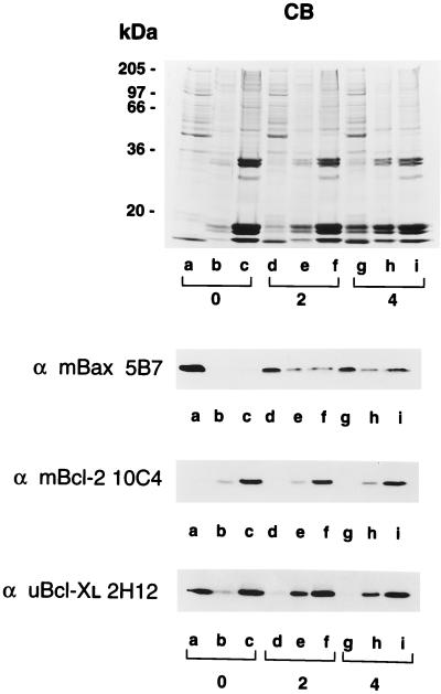
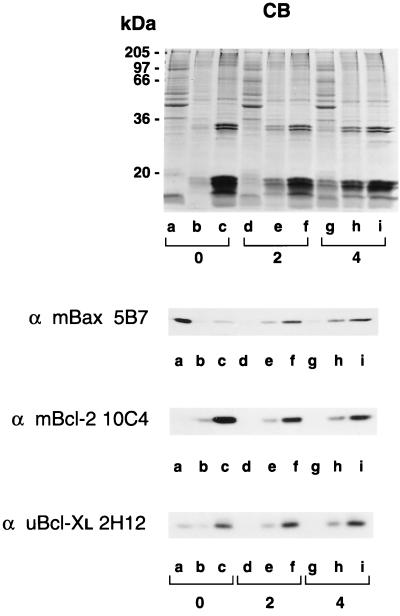
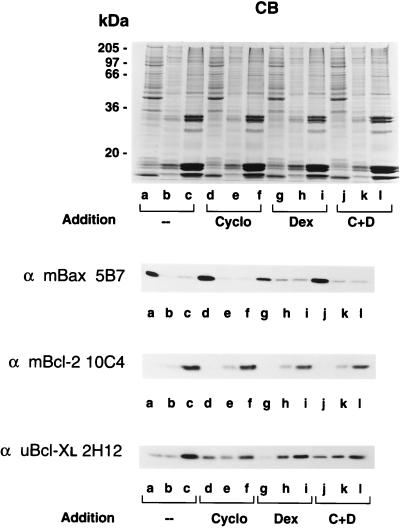
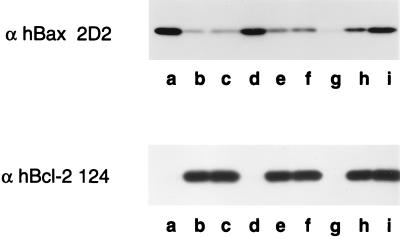
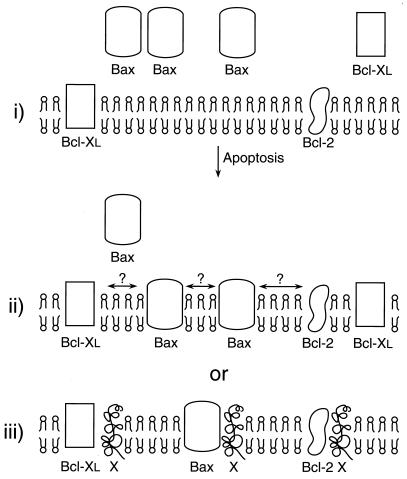
Bcl-2, Bcl-X(L), and Bax are members of the Bcl-2 family that play key roles in the regulation of apoptosis. These proteins are believed to be membrane bound and their ability to undergo both homodimerization and heterodimerization has been proposed to regulate apoptosis. Herein we report that in murine thymocytes, Bcl-2 is exclusively membrane-bound, whereas Bax is present predominantly in the cytosol and Bcl-X(L) is present in both soluble and membrane-bound forms. Induction of apoptosis in murine thymocytes by dexamethasone or gamma-irradiation shifts the subcellular locations of Bax and Bcl-X(L) from soluble to membrane-bound forms. A similar shift in the localization of Bax from the cytosol to membranes was observed in HL-60 leukemia cells upon induction of apoptosis by staurosporine. Inhibition of apoptosis with cycloheximide inhibits the movement of Bax and Bcl-X(L) in thymocytes from the cytosol into membranes induced by dexamethasone treatment. These movements may represent an important step in the pathway by which members of this family regulate apoptosis.


Figures

Figure 1
Figure 1
Differential localization of Bax and Bcl-XL and their association with membranes in response to dexamethasone-induced apoptosis. Apoptosis was induced in murine thymocytes by the addition of dexamethasone. Untreated cells (lanes a–c) and cells incubated 2 h (lanes d–f) and 4 h (lanes g–i) with dexamethasone were hypotonically lyzed, homogenized with a Dounce homogenizer, and separated into soluble (lanes a, d, and g), high-speed membrane pellet (lanes b, e, and h), and nuclear fractions (lanes c, f, and i) by differential centrifugation. The protein samples were analyzed by SDS/PAGE on gels stained with Coomassie blue (CB) or by Western blotting with anti-mBax 5B7, anti-mBcl-2 10C4, and anti-uBcl-XL 2H12 monoclonal antibodies.
Figure 2
Figure 2
Association of Bax and Bcl-XL with membranes in response to γ-irradiation-induced apoptosis. Apoptosis was induced in murine thymocytes by γ-irradiation. Untreated cells (lanes a–c) and cells collected 2 h (lanes d–f) and 4 h (lanes g–i) after irradiation were hypotonically lyzed, homogenized in a Dounce homogenizer, and separated into soluble (lanes a, d, and g), high-speed membrane pellet (lanes b, e, and h), and nuclear fractions (lanes c, f, and i) by differential centrifugation. The protein samples were analyzed by SDS/PAGE on gels stained with Coomassie blue (CB) or by Western blotting with anti-mBax 5B7, anti-mBcl-2 10C4, and anti-uBcl-XL 2H12 monoclonal antibodies.
Figure 3
Figure 3
Inhibition of dexamethasone-induced apoptosis by cycloheximide blocks migration of Bax and Bcl-XL into membranes. Murine thymocytes were incubated in the culture medium in the absence or in the presence of cycloheximide, dexamethasone, or a combination of cycloheximide and dexamethasone for 2 h. The cells were collected, hypotonically lyzed, homogenized in a Dounce homogenizer, and fractionated into soluble (lanes a, d, g, and j), high-speed membrane pellet (lanes b, e, h, and k), and nuclear fractions (lanes c, f, i, and l) by differential centrifugation. The samples were analyzed by SDS/PAGE on gels stained with Coomassie blue (CB) or by Western blotting with anti-mBax 5B7, anti-mBcl-2 10C4, and anti-uBcl-XL 2H12 monoclonal antibodies.
Figure 4
Figure 4
Staurosporine treatment of HL-60 cells promotes membrane association of Bax. HL-60 cells were treated either in absence (lanes a–c) or in the presence of 1 μM staurosporine for 2 h (lanes d–f) and 4 h (lanes g–i). The cells were collected, hypotonically lyzed, homogenized with a Dounce homogenizer, and fractionated into soluble (lanes a, d, and g), high-speed membrane pellet (lanes b, e, and h), and nuclear fractions (lanes c, f, and i) by differential centrifugation. The samples were analyzed by Western blotting with anti-hBax 2D2 and anti-hBcl-2 124 monoclonal antibodies.
Figure 5
Figure 5
Schematic representation of membrane insertion of Bax and Bcl-XL. In murine thymocytes, Bax is cytosolic, Bcl-2 is predominantly membrane bound, and Bcl-XL is present in both soluble and membrane-bound forms (i). Upon induction of apoptosis by dexamethasone or γ-irradiation, virtually all the cytosolic Bcl-XL and a significant amount of Bax becomes membrane-bound. It is not known whether Bax, in its membrane-associated state, undergoes hetero- or homodimerizations (ii). Alternatively, it is possible that Bax, Bcl-2, and Bcl-XL in their membrane-bound states may be competing for the binding of a yet unknown protein(s) in membranes to promote either cell death or survival (iii).

References

    1. Boise L H, Gottschalk A R, Quintáns J, Thompson C B. Curr Top Microbiol Immunol. 1995;200:107–121. - PubMed
    1. Reed J C. Hematol Oncol Clin North Am. 1995;9:451–473. - PubMed
    1. White E. Genes Dev. 1996;10:1–15. - PubMed
    1. Yang E, Korsmeyer S J. Blood. 1996;88:386–401. - PubMed
    1. Oltvai Z N, Milliman C L, Korsmeyer S J. Cell. 1993;74:609–619. - PubMed

Publication types