. 1997 Apr 15;94(8):3673-8.
doi: 10.1073/pnas.94.8.3673.

Buried asparagines determine the dimerization specificities of leucine zipper mutants

Affiliations

Buried asparagines determine the dimerization specificities of leucine zipper mutants

X Zeng et al. Proc Natl Acad Sci U S A. .

Abstract

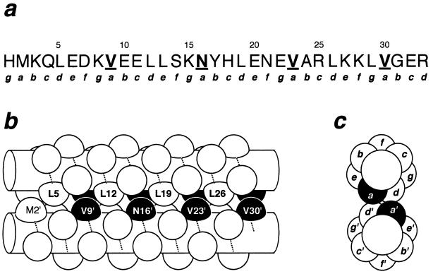
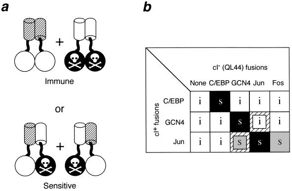
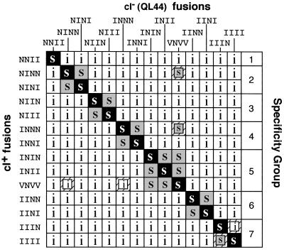
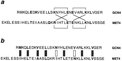
Regulation of gene expression by many transcription factors is controlled by specific combinations of homo- and heterodimers through a short alpha-helical coiled-coil known as a leucine zipper. The dimer interface of a leucine zipper involves side chains of the residues at the a, d, e, and g positions of the (abcdefg)n heptad repeat. To understand the basis for the specificity of dimer formation, we characterized GCN4 leucine zipper mutants with all 16 possible permutations and combinations of isoleucines and asparagines at four a positions in the dimer interface, using a genetic test for the specificity of dimer formation by lambda repressor-leucine zipper fusions. Heterodimers were detected by loss of repressor activity in the presence of a fusion to a dominant-negative mutant form of the DNA-binding domain of repressor. Reconstruction experiments using leucine zippers from GCN4, Jun, Fos, and C/EBP showed that this assay distinguishes pairs that form heterodimers from those that do not. We found that the mutants have novel dimerization specificities determined by the positioning of buried asparagine residues at the a positions. The pattern of buried polar residues could also explain the dimerization specificities of some naturally occurring leucine zippers. The altered specificity mutants described here should be useful for the construction of artificial regulatory circuitry.


Figures

Figure 1
Figure 1
The leucine zipper of GCN4 arranged as a parallel coiled coil. (a) Sequence of the leucine zipper as it occurs in the λ repressor fusion system. Lowercase letters indicate positions in the heptad repeat. The a positions targeted for mutagenesis are highlighted in boldface and are underlined. (b) Side view of the dimer. The amino acid backbones in a helical conformation are represented by cylinders with the path of the polypeptide chain indicated by the dotted lines. Side chains are represented as knobs. Residues targeted for mutagenesis are highlighted in black. (c) End view showing how different heptad positions are arranged in the dimer. The side chains at the a positions are buried in the dimer interface.
Figure 2
Figure 2
Rationale for a negative-dominance-based specificity assay with repressor fusions. (a) Active fusions (open circles fused to hatched cylinders) are coexpressed with fusions to an inactivated repressor domain (black circles marked by a skull and crossbones fused to open cylinders). If heterodimers do not form, cells will be immune to phage infection (Upper). Formation of heterodimers will titrate the active monomers into inactive heterodimers, and cells expressing these fusion proteins will be sensitive to killing by phage (Lower). (b) Reconstruction results with the leucine zippers of C/EBP, GCN4, Jun, and Fos. Pairwise combinations of fusion proteins with active (cI+) and inactive (cI/QL44) DNA-binding domains were expressed from compatible plasmids in E. coli. White boxes marked with an i indicate combinations that are immune to λ; s indicates combinations that are sensitive to λ. Black boxes indicate those pairs where the active and inactive DNA-binding domains are fused to the same leucine zipper; gray boxes indicate titration involving two different leucine zippers. Small inset boxes over a hatched background indicate one-way titrations.
Figure 3
Figure 3
Effects of a position mutations on dimerization specificity. A subset of the mutants shown in Table 1 were tested for heterodimer formation by the in vivo competition test shown in Fig. 1. Combinations that formed heterodimers and were sensitive to superinfection are indicated by an s. Combinations that retained immunity are indicated by an i. Shadings are as in Fig. 2. Groups of leucine zippers with overlapping specificities are indicated by brackets on the right.
Figure 4
Figure 4
Alignment of the leucine zippers of GCN4 and MET4. (a) Putative salt bridges involving the e and g positions (shown in boldface). Brackets indicate ion pairs observed in GCN4 (above the sequences) and predicted for MET4 (below) homodimers. Diagonal lines indicate similar ion pairs predicted to form in heterodimers. (b) Interactions in the hydrophobic core residues of putative heterodimers. The a and d positions are shown in boldface. Hatched boxes indicate interactions that would not discriminate between homodimers and heterodimers. Open boxes indicate mismatches between hydrophobic and polar side chains.

References

    1. O’Shea E K, Klemm J D, Kim P S, Alber T. Science. 1991;254:539–544. - PubMed
    1. Ellenberger T E, Brandl C J, Struhl K, Harrison S C. Cell. 1992;71:1223–1237. - PubMed
    1. Harbury P B, Zhang T, Kim P S, Alber T. Science. 1993;262:1401–1407. - PubMed
    1. Harbury P B, Kim P S, Alber T. Nature (London) 1994;371:80–83. - PubMed
    1. Glover J N M, Harrison S C. Nature (London) 1995;373:257–261. - PubMed

Publication types

LinkOut - more resources