. 1997 Apr 15;94(8):3795-800.
doi: 10.1073/pnas.94.8.3795.

Glucose transporter GLUT3: ontogeny, targeting, and role in the mouse blastocyst

Affiliations

Glucose transporter GLUT3: ontogeny, targeting, and role in the mouse blastocyst

M Pantaleon et al. Proc Natl Acad Sci U S A. .

Abstract

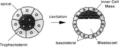
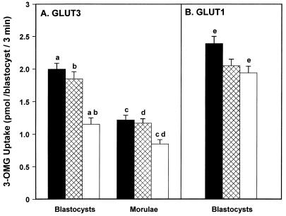
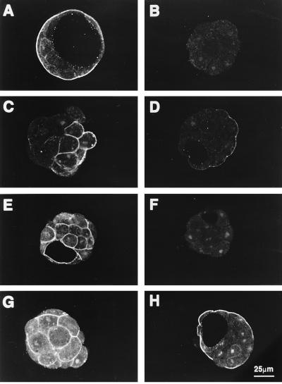
The first differentiative event in mammalian development is segregation of the inner cell mass and trophectoderm (TE) lineages. The epithelial TE cells pump fluid into the spherical blastocyst to form the blastocyst cavity. This activity is fuelled by glucose supplied through facilitative glucose transporters. However, the reported kinetic characteristics of blastocyst glucose transport are inconsistent with those of the previously identified transporters and suggested the presence of a high-affinity glucose carrier. We identified and localized the primary transporter in TE cells. It is glucose transporter GLUT3, previously described in the mouse as neuron-specific. In the differentiating embryo, GLUT3 is targeted to the apical membranes of the polarized cells of the compacted morula and then to the apical membranes of TE cells where it has access to maternal glucose. In contrast, GLUT1 was restricted to basolateral membranes of the outer TE cells in both compacted morulae and blastocysts. Using antisense oligodeoxynucleotides to specifically block protein expression, we confirmed that GLUT3 and not GLUT1 is the functional transporter for maternal glucose on the apical TE. More importantly, however, GLUT3 expression is required for blastocyst formation and hence this primary differentiation in mammalian development. This requirement is independent of its function as a glucose transporter because blastocysts will form in the absence of glucose. Thus the vectorial expression of GLUT3 into the apical membrane domains of the outer cells of the morula, which in turn form the TE cells of the blastocyst, is required for blastocyst formation.


Figures

Figure 1
Figure 1
Schematic representation of a compacted morula and blastocyst (adapted from ref. 1). Blastocyst formation is the morphological consequence of cavitation. During this process the newly differentiated epithelial TE cells, with their apical surface facing outward, engage in vectorial fluid transport so that the resultant blastocyst consists of TE cells surrounding a fluid filled cavity (blastocoel) and a small group of totipotent inner cell mass cells eccentrically placed at the side of one pole of the TE.
Figure 2
Figure 2
Expression of a 284-bp fragment corresponding to murine GLUT3 mRNA in early mouse embryos. Each embryo lane was produced using a cDNA sample derived from RNA of 10 embryos. Brain and 3T3 fibroblast cell lanes were produced using cDNA derived from ≈20 ng RNA. The RNA preparations were reverse transcribed and amplified by 40 cycles of PCR as described. Lanes: L, DNA ladder (bands from top to bottom: 603, 303, 291/281, 234, and 194 bp); 1, negative control (no cDNA); 2, brain; 3, 3T3 fibroblasts; 4, fertilized eggs; 5, two-cell embryos; 6, four-cell embryos; 7, morulae; 8, blastocysts.
Figure 3
Figure 3
Cellular localization of GLUT3 and GLUT1 during mouse preimplantation development. Confocal images of optical sections of early four-cell, six-cell embryos, early morula, compacted morula, and blastocyst incubated with 10 μg/ml anti-GLUT3 antibody (BF); morula and blastocyst incubated with anti-GLUT1 antibody (G and H); and blastocyst incubated with pooled nonimmune rabbit serum (A). GLUT3 protein is first apparent at the late four- to six-cell stage (C) when it appears restricted to cytoplasmic vesicles. Note that, as the embryo develops, positive immunoreactivity first appears on plasma membranes then concentrates in the apical surface of the polarizing outer cells of the compacted morula. In blastocysts it is restricted to the apical membranes of TE. This contrasts with GLUT1 expression which is localized in basolateral membranes of both morulae (G) and blastocysts (H).
Figure 7
Figure 7
Immunoblot of GLUT3 expression in embryos following antisense oligodeoxynucleotide treatment. Groups of 80 two-cell embryos were cultured over 48 hr in BMOC2 (lane 2) or BMOC2 supplemented with 30 μM of either GLUT3 sense (lane 3) or GLUT3 antisense (lane 4). Resultant embryos were lysed, electrophoresed, and blotted with anti-GLUT3 antibody as described. Rat brain (lane 1), included as a positive control for GLUT3, showed a band at about 55 kDa which was also present in embryos from all treatments. GLUT3 immunoreactivity, however, was markedly reduced in antisense-treated embryos relative to control and sense treatments.
Figure 4
Figure 4
Effect of GLUT3 or GLUT1 antisense oligodeoxynucleotide treatment on blastocyst formation. Development of two-cell embryos cultured for 48 hr in BMOC2 (▪; control) supplemented with either 30 μM sense (⊠) or antisense (□) oligonucleotides for GLUT1 or GLUT3. Each bar represents the percentage of embryos forming blastocysts from three different experiments, each with a minimum of 20 embryos in each of the three treatments (i.e., 162, 256, and 178 embryos in control, antisense, and sense treatments, respectively). χ2 analyses showed a significant difference between treatments for the GLUT3 experiments only (∗∗∗, P < 0.0001). These GLUT3 data were further analyzed using three 2 × 2 contingency tables and individual treatments compared (control vs. sense, χ2 = 1.1452, P = 0.2846; control vs. antisense, χ2 = 29.299, P < 0.0001; sense vs. antisense, χ2 = 18.145, P < 0.0001).
Figure 5
Figure 5
Effect of GLUT3 (A) and GLUT1 (B) antisense oligodeoxynucleotide treatment during development on embryonic glucose transport activity. Uptake of 25 mM [3H]3-OMG over 3 min at 37°C in either resultant morulae or blastocysts following 48 hr culture in BMOC2 medium (▪; control) supplemented with either 30 μM sense (⊠) or antisense (□) oligodeoxynucleotides. Each bar represents the mean ± SEM of three experiments each containing three to six embryos in each treatment. Means with the same superscript are significantly different by ANOVA: a, b, c, d, P < 0.01; e, P < 0.05. Note that in GLUT1 experiments (B), transport activity of antisense-treated embryos is not significantly different from sense-treated embryos.
Figure 6
Figure 6
GLUT3 and GLUT1 immunofluorescence following antisense oligodeoxynucleotide treatment. Two-cell embryos developing over 48 hr in BMOC2 and supplemented with 30 μM oligodeoxynucleotides: GLUT3 sense (A), GLUT3 antisense (B, C, D, and G), GLUT1 sense (E), GLUT1 antisense (F and H); and then probed with anti-GLUT3 (AD and H) or anti-GLUT1 (EG) as described. GLUT3 immunoreactivity present in sense (A) cultured embryos is abolished by GLUT3 antisense treatment (B). Highly attenuated but not fully abolished GLUT3 immunoreactivity was observed in 10% of morulae (C) and all blastocysts examined (D). The specificity of both the antibodies and the oligonucleotide treatments is shown by the persistence of GLUT1 immunoreactivity in the GLUT3 antisense-treated embryos (G) and GLUT3 immunoreactivity following GLUT1 antisense treatment (H).

References

    1. Fleming T P. In: Epithelial Organization and Development. 1st Ed. Fleming T P, editor. London: Chapman & Hall; 1992. pp. 111–130.
    1. Leese H J, Barton A M. J Reprod Fertil. 1984;72:9–13. - PubMed
    1. Gardner D K, Leese H J. Development (Cambridge, UK) 1988;104:423–429. - PubMed
    1. Chi M M, Manchester J K, Basuray R, Mahendra S, Strickler R C, McDougal D B, Jr, Lowry O H. Proc Natl Acad Sci USA. 1993;90:10023–10025. - PMC - PubMed
    1. Manezwala F M, Cragoe E J, Jr, Schultz R M. Dev Biol. 1989;133:210–220. - PubMed

Publication types

Substances

LinkOut - more resources