. 1997 Apr 15;94(8):3811-6.
doi: 10.1073/pnas.94.8.3811.

Evolution of antifreeze glycoprotein gene from a trypsinogen gene in Antarctic notothenioid fish

Affiliations

Evolution of antifreeze glycoprotein gene from a trypsinogen gene in Antarctic notothenioid fish

L Chen et al. Proc Natl Acad Sci U S A. .

Abstract

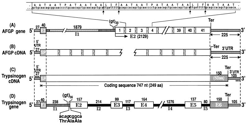
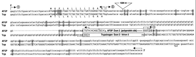
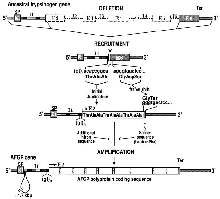
Freezing avoidance conferred by different types of antifreeze proteins in various polar and subpolar fishes represents a remarkable example of cold adaptation, but how these unique proteins arose is unknown. We have found that the antifreeze glycoproteins (AFGPs) of the predominant Antarctic fish taxon, the notothenioids, evolved from a pancreatic trypsinogen. We have determined the likely evolutionary process by which this occurred through characterization and analyses of notothenioid AFGP and trypsinogen genes. The primordial AFGP gene apparently arose through recruitment of the 5' and 3' ends of an ancestral trypsinogen gene, which provided the secretory signal and the 3' untranslated region, respectively, plus de novo amplification of a 9-nt Thr-Ala-Ala coding element from the trypsinogen progenitor to create a new protein coding region for the repetitive tripeptide backbone of the antifreeze protein. The small sequence divergence (4-7%) between notothenioid AFGP and trypsinogen genes indicates that the transformation of the proteinase gene into the novel ice-binding protein gene occurred quite recently, about 5-14 million years ago (mya), which is highly consistent with the estimated times of the freezing of the Antarctic Ocean at 10-14 mya, and of the main phyletic divergence of the AFGP-bearing notothenioid families at 7-15 mya. The notothenioid trypsinogen to AFGP conversion is the first clear example of how an old protein gene spawned a new gene for an entirely new protein with a new function. It also represents a rare instance in which protein evolution, organismal adaptation, and environmental conditions can be linked directly.


Figures

Figure 1
Figure 1
Structures of AFGP and trypsinogen genes and cDNAs from Antarctic notothenioid fish D. mawsoni. (A) AFGP gene in genomic clone Dm1A. The AFGP polyprotein coding region (2129 nt) contains 41 copies of AFGP coding sequences (boxed numbers) tandemly linked by highly conserved 9-nt spacers (bars filled with zigzagged lines). Nucleotide and translated amino acid sequence covering copies 2, 3, and (partly) 4 (extended above gene structure) is given. Posttranslational cleavage (↑ ↑) of spacers produces the mature AFGPs. Exon–intron boundaries were determined after two partial AFGP cDNAs were characterized (B). 5′ UTR and signal peptide (SP) sequence are encoded by E1, and AFGP polyprotein by E2. The single, intervening intron I1 is 1879 nt. (C) Trypsinogen cDNA contains a 27-nt 5′ UTR, 747 nt of pretrypsinogen coding sequence, and a 105-nt 3′ UTR. (D) Trypsinogen gene contains six exons (E1-E6) and five introns (I1-I5) with lengths in nt as indicated. Three regions of sequence identities are found on alignment of all four structures. The two pairs of dashed lines delimit the 5′ (67-nt) and 3′ (225-nt) end regions of sequence identities (gray regions) with the segments of the gene or encoded protein each represents, as labeled. The third region of identity is indicated by striped bars; I1 of trypsinogen gene (D) is found in two segments in I1 of AFGP gene (A) and both introns contain repetitive (gt)n at its 3′ end. The 9-nt element in trypsinogen gene that translates into the repeat unit of AFGP peptide backbone, Thr-Ala-Ala, straddles I1 and E2 (splice sequence in italics), as shown in D. Asterisks in C (trypsinogen cDNA) indicate primer sequences cTryp-AF5′ and cTryp-AF3′ used for RT-PCR of AFGP cDNA and trypsinogen gene. Structures are not necessarily drawn to scale.
Figure 2
Figure 2
Alignment of D. mawsoni AFGP and trypsinogen (Tryp) genomic sequences showing the three regions of high sequence identity between the two genes. ①, 5′ UTR (lowercase) and signal peptide coding sequences (uppercase; translated amino acids also shown) are 94% identical; ②, intron I sequences (lowercase) are 93% identical (position of the extra 1684-nt AFGP intron sequence is indicated); and ③, AFGP penultimate codon plus 3′UTR sequence (lowercase) is 96% identical to trypsinogen exon 6 (uppercase) and 3′ UTR (lowercase). Positions of gene-specific sequences are boxed. The small number of nucleotide differences in the three regions are highlighted. The dots underscore the 9-nt Thr-Ala-Ala coding element (acagcggca) in trypsinogen that might have been amplified to give rise to the repetitive tripeptide coding sequence of AFGP (in parentheses).
Figure 3
Figure 3
Southern blot of products from PCR-amplification of genomic DNA from three different notothenioids, using common 5′ and 3′ trypsinogen/AFGP primers (sites indicated by asterisks in Fig. 1C) and hybridized to an AFGP-specific probe containing the repetitive AFGP coding sequence only. The multiple AFGP-positive bands indicate that members of notothenioid AFGP gene families all have the hybrid AFGP/trypsinogen gene structure. Dm, Dissostichus mawsoni; Nc, Notothenia coriiceps; Pb, Pagothenia borchgrevinki.
Figure 4
Figure 4
Likely mechanism by which an ancestral trypsinogen gene was transformed into an AFGP gene. The 5′ end (E1, I1, and small segment of E2) and the 3′ end (I5 3′ splice site and E6) of trypsinogen gene were recruited and linked, and the remainder of the gene deleted (dashed lines and boxes). The Thr-Ala-Ala coding element was duplicated, presumably via slippage at the repetitive (gt)n sequence during replication. The recruited E1 provided the 5′ UTR and signal peptide sequences for the new AFGP gene. The deletion, linking, and amplification events led to a 1-nt frameshift resulting in a termination codon (tga) at the start of the recruited trypsinogen E6 and converting it into the 3′ flanking sequence of the AFGP gene. The spacer sequence (bars filled with zigzagged lines) and additional I1 sequence might be existing sequence in the trypsinogen progenitor gene or acquired through recombinatory events. The Thr-Ala-Ala coding duplicants plus a spacer became amplified de novo to form the new AFGP polyprotein coding region. The regions of identity are illustrated as in Fig. 1. Splice sites in trypsinogen gene are given in italics.

References

    1. Gon O, Heemstra P C. Fishes of the Southern Ocean. Grahamstown, South Africa: JLB Smith Institute of Ichthyology; 1990.
    1. Hubold G. In: Biology of Antarctic Fish. di Prisco G, Maresca M, Tota B, editors. Berlin: Springer; 1991. pp. 3–22.
    1. Eastman J T. Antarctic Fish Biology: Evolution in a Unique Environment. San Diego: Academic; 1993.
    1. DeWitt H H. In: Antarctic Map Folio Series, Folio 15. Bushnell V C, editor. New York: Am. Geogr. Soc.; 1971. pp. 1–10.
    1. Ekau W. Antarct Sci. 1990;2:129–137.

Publication types