. 1997 Apr 15;94(8):3943-7.
doi: 10.1073/pnas.94.8.3943.

Transgenic expression of CD95 ligand on islet beta cells induces a granulocytic infiltration but does not confer immune privilege upon islet allografts

Affiliations

Transgenic expression of CD95 ligand on islet beta cells induces a granulocytic infiltration but does not confer immune privilege upon islet allografts

J Allison et al. Proc Natl Acad Sci U S A. .

Abstract

Binding of CD95 (Fas/APO-1) by its ligand (CD95L) commonly induces apoptosis. Apoptosis of activated T cells, induced by CD95L expressed in the rodent testis, has been proposed to be the mechanism of immune privilege [Bellgrau, D., Gold, D., Selawry, H., Moore, J., Franzusoff, A. & Duke, R. C. (1995) Nature (London) 377, 630-632]. To test whether CD95L could protect pancreatic islet grafts from rejection, we made transgenic mice expressing murine CD95L on their islet beta cells and transplanted fetal pancreata under the kidney capsules of allogeneic animals. Expression of CD95L failed to protect the grafts from rejection. However, transgenic mice developed a granulocytic infiltration in their pancreata. These results demonstrate a pro-inflammatory function of CD95L and suggest that expression of CD95L may not be sufficient to protect organ allografts.
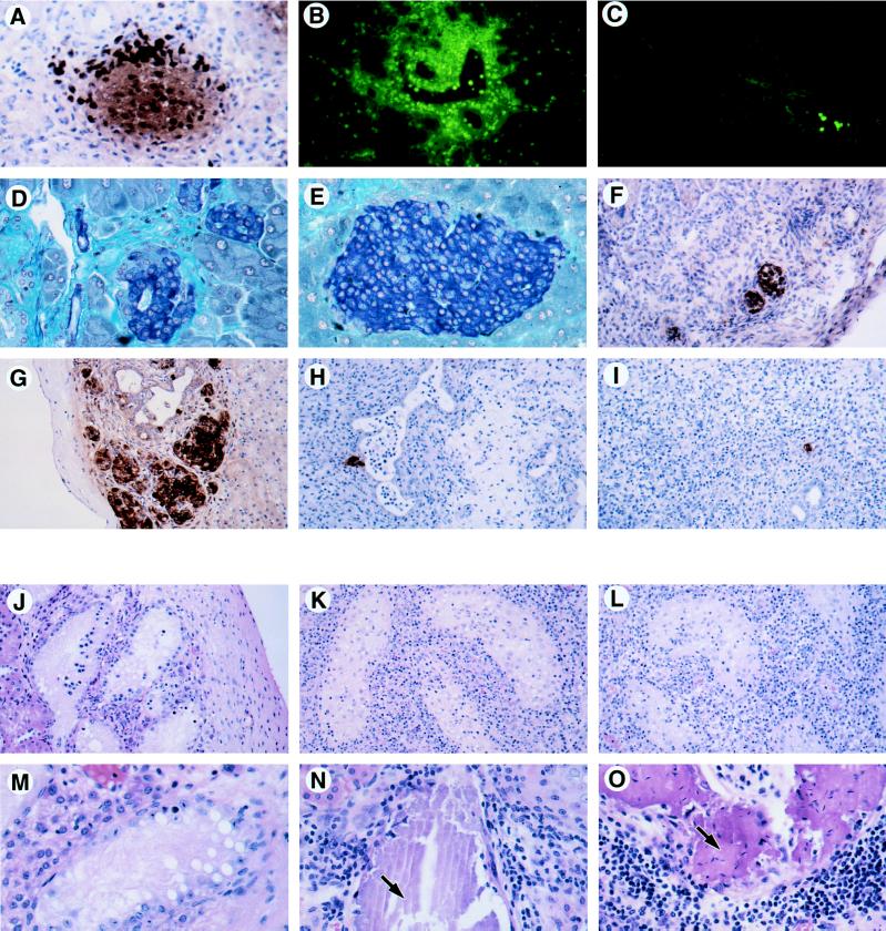

Figures

Figure 1
Figure 1
Expression of CD95L by transgenic islet β cells (A) and by Sertoli cells in the testis (B). Cryostat sections from a RIP–CD95Lhi pancreas or a normal testis were fixed in acetone and then stained for CD95L using a rabbit polyclonal antibody detected with an fluorescein isothiocyanate-coupled antibody to rabbit Ig. The same tissues were stained with an irrelevant rabbit polyclonal antibody (C and D).
Figure 2
Figure 2
Killing of SKW6 B lymphoblastoid cells by anti-CD95 antibody or by RIP–CD95L+ adult islet β cells. SKW6 B cells are killed by increasing concentrations of anti-CD95 antibody (left side of graph) or by incubation with RIP–CD95L+ adult islet cells expressing different amounts of transgenic CD95L (right side of graph). A CD95–Fcγ fusion protein that binds to and blocks CD95L was added where indicated. Results shown are averages of three experiments performed on separate occasions. Error bars indicate 2 × SEM.
Figure 3
Figure 3
Pathology of in situ and transplanted CD95L+ tissue. (A) Insulin staining by immunoperoxidase (brown cells) of a RIP–CD95Lhi islet from a 12-day-old mouse pancreas. The very dark stained cells are granulocytes (neutrophils and eosinophils) expressing endogenous peroxidase. (×200.) (B and C) Gr-1 staining (green cells) of a RIP–CD95Lhi 8-week-old pancreas (B) and a nontransgenic control pancreas (C). Gr-1+ neutrophils infiltrate the transgenic pancreas but are only seen in a vessel of the nontransgenic pancreas. (B and C, ×100.) (D and E) Gomori’s aldehyde fuchsin staining for insulin (blue cells) of an adult RIP–CD95Lhi islet (D) and a nontransgenic control islet (E). The small disorganized islets were typical of the RIP–CD95Lhi adult transgenic pancreas and contained only a few infiltrating granulocytes and macrophages. (D and E, ×200.) (F) CD95L staining (brown cells) of a RIP–CD95Lhi syngeneic (B6 to B6) islet graft after 14 days. (×100.) (G) Insulin expression (brown cells) in a nontransgenic control islet graft (B6 to B6) after 14 days. (×100.) (H and I) Insulin staining cells in allogeneic islet grafts (B6 to BALB/c) from a RIP-CD95L donor (H) or a nontransgenic donor (I) after 14 days. (×100.) (JO) Hematoxylin/eosin staining of testis grafts recovered from BALB/c male recipients after 14 days (JL) and after 28 days (MO). (JL, ×100; MO, ×200.) (J and M) Syngeneic grafts (BALB/c). (K) Allogeneic graft (C3H). (L and O) Allogeneic grafts (C3Hgld). (N) Allogeneic graft (B6). Arrows in N and O point to regions of calcification. Sections in AC and F were acetone-fixed frozen sections; sections in D, E, and GO were Bouin’s solution-fixed paraffin sections.

Comment in

  • Fas-ligand: privilege and peril.
    Green DR, Ware CF. Green DR, et al. Proc Natl Acad Sci U S A. 1997 Jun 10;94(12):5986-90. doi: 10.1073/pnas.94.12.5986. Proc Natl Acad Sci U S A. 1997. PMID: 9177153 Free PMC article. Review. No abstract available.

References

    1. Bellgrau D, Gold D, Selawry H, Moore J, Franzusoff A, Duke R C. Nature (London) 1995;377:630–632. - PubMed
    1. Griffith T S, Brunner T, Fletcher S M, Green D R, Ferguson T A. Science. 1995;270:1189–1192. - PubMed
    1. Barker C F, Billingham R E. Adv Immunol. 1977;25:1–54. - PubMed
    1. Nagata S, Golstein P. Science. 1995;267:1449–1456. - PubMed
    1. Klas C, Debatin K M, Jonker R R, Krammer P H. Int Immunol. 1993;5:625–630. - PubMed

Publication types