

N-terminal sequences from Autographa californica nuclear polyhedrosis virus envelope proteins ODV-E66 and ODV-E25 are sufficient to direct reporter proteins to the nuclear envelope, intranuclear microvesicles and the envelope of occlusion derived virus
- PMID: 9108103
- PMCID: PMC20566
- DOI: 10.1073/pnas.94.8.4050
N-terminal sequences from Autographa californica nuclear polyhedrosis virus envelope proteins ODV-E66 and ODV-E25 are sufficient to direct reporter proteins to the nuclear envelope, intranuclear microvesicles and the envelope of occlusion derived virus
Abstract
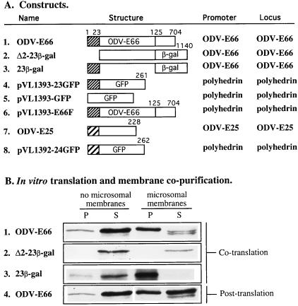
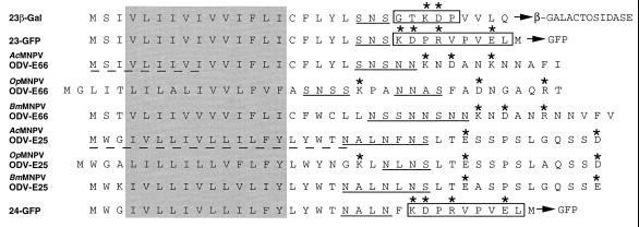
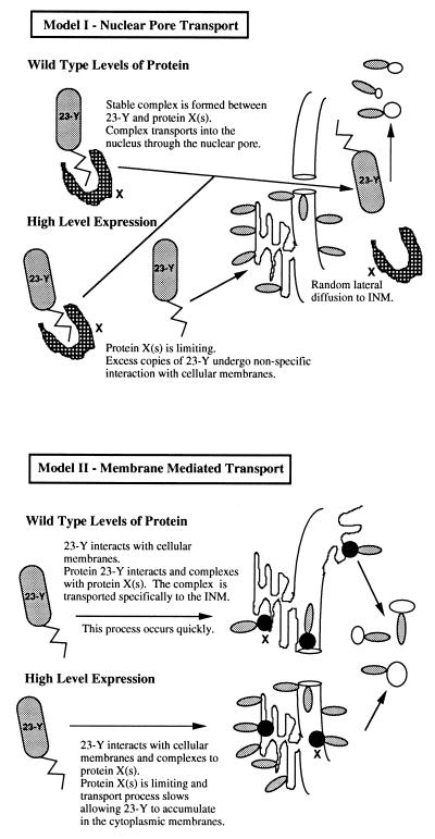
Baculovirus occlusion-derived virus (ODV) derives its envelope from an intranuclear membrane source. N-terminal amino acid sequences of the Autographa californica nuclear polyhedrosis virus (AcMNPV) envelope proteins, ODV-E66 and ODV-E25 (23 and 24 amino acids, respectively) are highly hydrophobic. Recombinant viruses that express the two N-terminal amino acid sequences fused to green fluorescent protein (23GFP or 24GFP) provided visual markers to follow protein transport and localization within the nucleus during infection. Autoflourescence was first detected along the cytoplasmic periphery of the nucleus and subsequently localized as foci to discrete locations within the nucleus. Immunoelectron microscopy confirmed that these foci predominantly contained intranuclear microvesicles and the reporter fusion proteins were also detected in cytoplasmic membranes near the nucleus, and the outer and inner nuclear membrane. Therefore, these defined hydrophobic domains are sufficient to direct native and fusion proteins to induced membrane microvesicles within a baculovirus-infected cell nucleus and the viral envelope. In addition, these data suggest that movement of these proteins into the nuclear envelope may initiate through cytoplasmic membranes, such as endoplasmic reticulum, and that transport into the nucleus may be mediated through the outer and inner nuclear membrane.
Figures
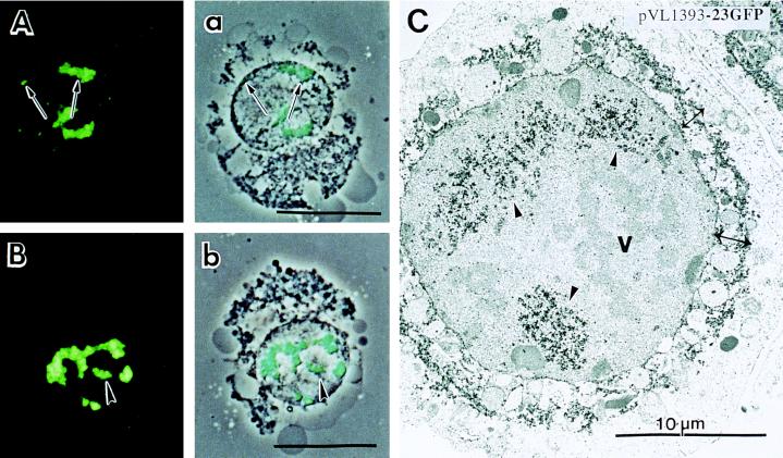
 ), Intranuclear microvesicles, (↔), cytoplasmic
membranes condensed near the nuclear envelope. Protein was detected as
in C. C, cytoplasm; n,
nucleus.
), Intranuclear microvesicles, (↔), cytoplasmic
membranes condensed near the nuclear envelope. Protein was detected as
in C. C, cytoplasm; n,
nucleus.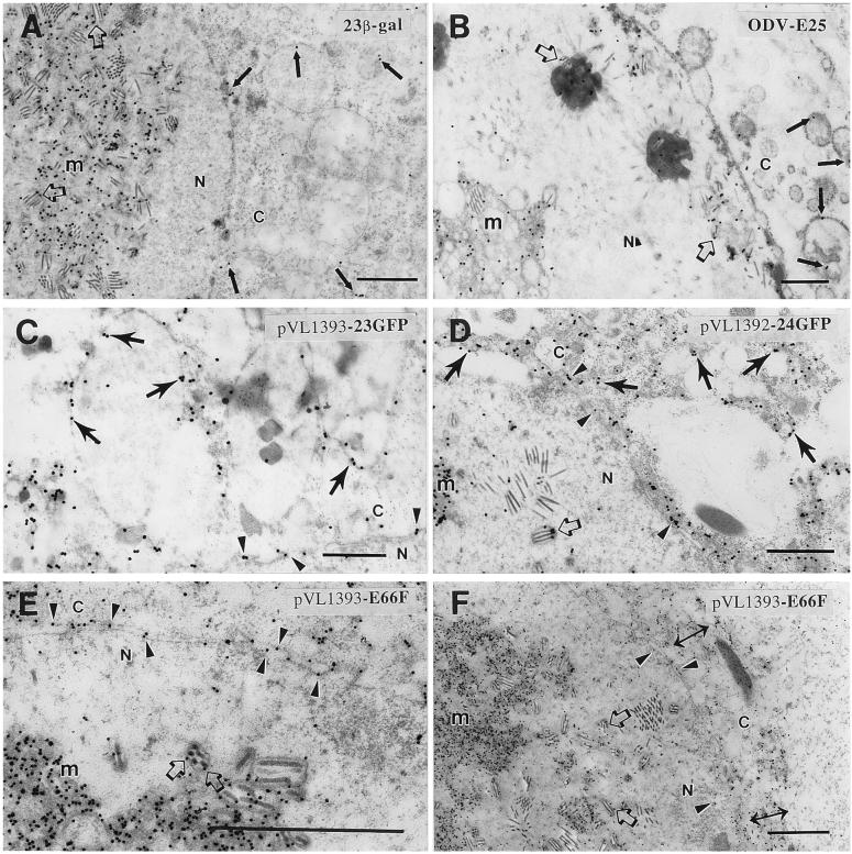
References
-
- Wiley D C, Skehel J J. In: Virology. Fields B N, Knipe D M, Chanock R M, Hirsch M S, Milnick J L, Monath T P, Roizman B, editors. New York: Raven; 1990. pp. 63–85.
-
- Adams J R, McClintock J T. In: Atlas of Invertebrate Viruses. Adams J R, Bonami J R, editors. Boca Raton, FL: CRC; 1991. pp. 87–226.
-
- Blissard G W, Rohrmann G F. Annu Rev Entomol. 1990;35:127–155. - PubMed
-
- Fraser M J. J Ultrastruct Mol Struct Res. 1986;95:189–195.
-
- Hong T, Braunagel S C, Summers M D. Virology. 1994;204:210–222. - PubMed
Publication types
MeSH terms
Substances
Grants and funding
LinkOut - more resources
Full Text Sources
Other Literature Sources
