. 1997 Apr 15;94(8):4056-61.
doi: 10.1073/pnas.94.8.4056.

A diffusible cytotoxin of Haemophilus ducreyi

Affiliations
Free PMC article

A diffusible cytotoxin of Haemophilus ducreyi

L D Cope et al. Proc Natl Acad Sci U S A. .
Free PMC article

Abstract

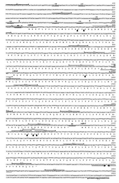
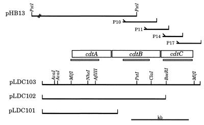
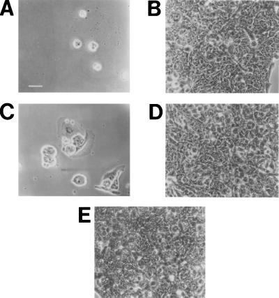
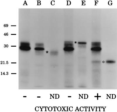
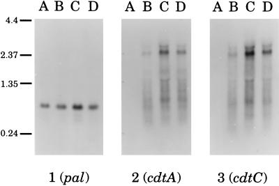
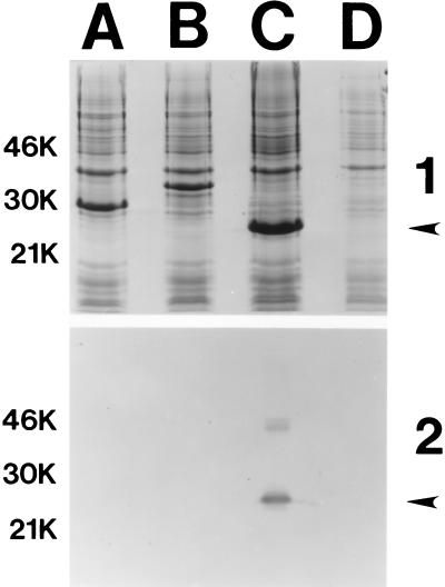
Little is known about the virulence mechanisms employed by Haemophilus ducreyi in the production of genital ulcers. This Gram-negative bacterium previously has been shown to produce a soluble cytotoxic activity that kills HeLa and HEp-2 cells. We have now identified a cluster of three H. ducreyi genes that encode this cytotoxic activity. The predicted proteins encoded by these genes are most similar to the products of the Escherichia coli cdtABC genes that comprise the cytolethal distending toxin (CDT) of this enteric pathogen. Eleven of 12 H. ducreyi strains were shown to possess this gene cluster and culture supernatants from these strains readily killed HeLa cells. The culture supernatant from a single strain of H. ducreyi that lacked these genes was unable to kill HeLa cells. When the H. ducreyi cdtABC gene cluster was cloned into E. coli, culture supernatant from the recombinant E. coli clone killed HeLa cells. A monoclonal antibody that neutralized this soluble cytotoxic activity of H. ducreyi was shown to bind to the H. ducreyi cdtC gene product. This soluble H. ducreyi cytotoxin may play a role in the development or persistence of the ulcerative lesions characteristic of chancroid.


Figures

Figure 2
Figure 2
Nucleotide sequence of the H. ducreyi strain 35000 cdtABC genes together with the deduced amino acid sequence of the predicted proteins. Putative −35 and −10 regions are underlined, as are the putative ribosomal binding sites (SD). An inverted repeat located 3′ from the end of the cdtC gene is indicated by →← opposing arrows. Oligonucleotide primers (P) used in PCR reactions are numbered above the relevant sequences. Putative signal peptidase I cleavage sites are indicted by ↑. Putative transcription initiation sites detected by primer extension studies are indicated by ∗.
Figure 1
Figure 1
Partial restriction enzyme map of the H. ducreyi cdtABC gene cluster. The 11-kb insert from the recombinant plasmid pHB13 is shown in truncated form. The relative position of the four oligonucleotide primers (P10, P11, P14, and P17) used in ligation-based PCR reactions (together with the T3 or T7 primers) to obtain initial sequence information for the downstream half of the cdtB gene and all of the cdtC gene are indicated together with the size of the relevant PCR product. The thin shaded bars (░⃞) beneath the ORFs demarcate the probes used for both Northern and Southern blot analyses. Plasmids pLDC101, pLDC102, and pLDC103 contain the H. ducreyi cdtA, cdtAB, and cdtABC genes as indicated.
Figure 3
Figure 3
Killing of HeLa cells by wild-type and recombinant H. ducreyi cdt gene products. Partial monolayers of HeLa cells (20–30% confluent) in 24-well tissue culture plates were exposed to culture supernatants (A–D) or to bacterial culture medium (E) for 2 hr. The wells were stained and photographed 72 hr later. (A) H. ducreyi strain 35000; (B) H. ducreyi strain 512; (C) E. coli DH5α(pLDC103); (D) E. coli DH5α(pCRII); (E) Luria–Bertani culture medium. (Bar = 50 μm.)
Figure 4
Figure 4
Fluorographic detection of H. ducreyi CDT proteins expressed in vitro. The H. ducreyi cdtA gene (lane C), the H. ducreyi cdtB gene (lane E), and the H. ducreyi cdtC gene (lane G) were each amplified from the chromosome of H. ducreyi strain 35000 and used in an in vitro coupled transcription/translation system for linear DNA templates to establish the size of the protein product of each gene. The position of CdtA is indicated by ∗, that of CdtB by ▪, and that of CdtC by ○. The vector pCRII (lane A) and recombinant plasmids containing the H. ducreyi cdtA gene (pLDC101, lane B), the H. ducreyi cdtAB gene pair (pLDC102, lane D), and the entire H. ducreyi cdtABC gene cluster (pLDC103, lane F) were used in an in vitro coupled transcription/translation system for circular DNA templates. The HeLa cell killing ability (cytotoxic activity) of culture supernatants from E. coli DH5α-derived recombinant strains containing pLDC101, pLDC102, pLDC103, and the vector plasmid pCRII is indicated. ND, not determined.
Figure 5
Figure 5
Northern blot analysis of transcripts from the H. ducreyi cdt and pal genes. Total RNA prepared from cells of strain 512 (lane A), 35000 (lane B), RO18 (lane C), and STD102 (lane D) was analyzed with a pal-specific probe (panel 1), a cdtA-specific probe (panel 2), and a cdtC-specific probe (panel 3). Size markers (in kb) are shown on the left side of this figure.
Figure 6
Figure 6
Western blot-based identification of the H. ducreyi cdt gene product bound by the H. ducreyi cytotoxin-neutralizing mAb. After isopropyl β-d-thiogalactoside induction, proteins in recombinant E. coli strains carrying genes encoding the His-CDT fusion proteins were resolved by SDS/PAGE and either stained with Coomassie blue (panel 1) or probed in Western blot analysis with the cytotoxin-neutralizing mAb (panel 2). Lanes: A, His-CdtA; B, His-CdtB; C, His-CdtC; D, vector only. The arrows on the right side of each panel indicate the position of the 22-kDa His-CdtC fusion protein that bound the cytotoxin-neutralizing mAb. Size markers (in kDa) are present on the left side of each panel.

Similar articles

Cited by

References

    1. Trees D L, Morse S A. Clin Microbiol Rev. 1995;8:357–375. - PMC - PubMed
    1. Ronald A R, Albritton W L. In: Sexually Transmitted Diseases. Holmes K K, Mardh P-A, Sparling P F, Wiesner P J, editors. New York: McGraw–Hill; 1990. pp. 263–271.
    1. Cameron D W, Simonsen J N, D’Costa L J, Ronald A R, Maitha G M, Gakinya M N, Cheang M, Piot P, Brunham R C, Plummer F A. Lancet. 1989;ii:403–408. - PubMed
    1. Plummer F A, Wainberg M A, Plourde P, Jessamine P G, D’Costa J L, Wamola I A, Ronald A R. J Infect Dis. 1990;161:810–811. - PubMed
    1. Odumeru J A, Wiseman G M, Ronald A R. J Med Microbiol. 1987;23:155–162. - PubMed

Publication types

Substances

Associated data

LinkOut - more resources