

Review
doi: 10.1016/s0966-842x(97)84657-4.
Cell invasion by the vertebrate stages of Plasmodium
Affiliations
- PMID: 9108930
- PMCID: PMC5538855
- DOI: 10.1016/s0966-842x(97)84657-4
Item in Clipboard
Review
Cell invasion by the vertebrate stages of Plasmodium
Trends Microbiol.
1997 Feb.
Abstract
Protozoans of the genus Plasmodium are the causative agents of malaria; they have a complex life cycle involving vertebrate and arthropod hosts and have three distinct invasive stages. Although the invasive stages probably invade cells using similar mechanisms, each stage has a different host cell specificity and utilizes different receptors to enter cells.
Figures
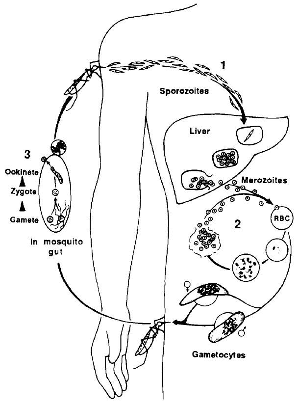
Life cycle of the malaria parasite. (1) Sporozoites are injected into a vertebrate host during the blood meal of a female anopheline mosquito and rapidly invade hepatocytes. One sporozoite can develop into 20 000 merozoites, which rupture from the hepatocyte, enter the bloodstream and invade erythrocytes. (2) In the asexual erythrocytic cycle, merozoites invade erythrocytes and mature from ring stages to schizonts within these cells in 48–72 h, the time varying with the species of malaria parasite. (3) Some erythrocytic stages differentiate into gametocytes, which are infective for mosquitoes. Fertilization occurs in the mosquito midgut, and within 24 h zygotes transform into ookinetes, which penetrate the midgut and form oocysts. Sporozoites rupture from these oocysts and invade the salivary glands of the mosquito from where they are injected into a vertebrate host.
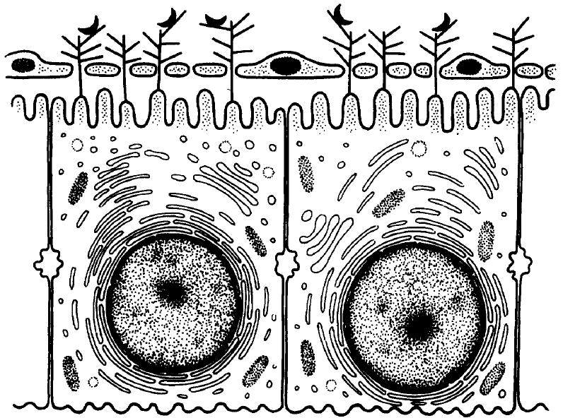
Diagrammatic representation of direct interaction between sporozoites and hepatocytes. Heparan sulfate proteoglycans (HSPGs) are shown as branched structures on the hepatocyte membrane, protruding through the open fenestrae of the endothelial cells into the sinusoidal lumen. Sporozoites, shown as black half-moon structures, are captured by these HSPGs and then invade hepatocytes.
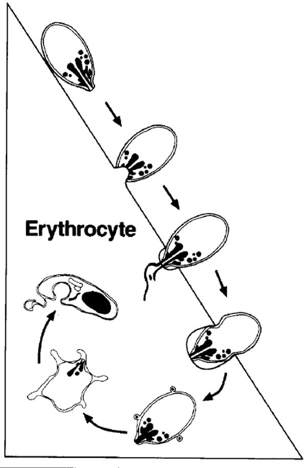
Schematic representation of the major morphological events of merozoite invasion. On top, the merozoite is depicted in the attachment and orientation phase, followed by binding, junction formation and rhoptry discharge. The merozoite is then shown entering the parasitophorous vacuole as the junction between the merozoite and the erythrocyte moves towards the posterior end of the merozoite. The parasite is then completely intracellular and flattens with its cytoplasm remaining thick only at the edges, giving the appearance of a ‘ring’.
References
-
- Vanderberg J, Chew S, Stewart MJ. J Protozool. 1990;37:528–536. - PubMed
-
- Ward G, Chitnis CE, Miller LH. In: Strategtes for Intracellular Survival of Microbes. Russell D, editor. Saunders; 1994. pp. 155–190.
-
- Ungureanu E, et al. Trans R Soc Trop Med Hyg. 1976;70:482–483. - PubMed
-
- Ponnudurai T, et al. Trans R Soc Trop Med Hyg. 1991;85:175–180. - PubMed
Publication types
MeSH terms
Substances
Grants and funding
LinkOut - more resources
Full Text Sources
Other Literature Sources
