Comparative Study
. 1997 Apr 29;94(9):4330-5.
doi: 10.1073/pnas.94.9.4330.

P110delta, a novel phosphoinositide 3-kinase in leukocytes

Affiliations
Comparative Study

P110delta, a novel phosphoinositide 3-kinase in leukocytes

B Vanhaesebroeck et al. Proc Natl Acad Sci U S A. .

Abstract

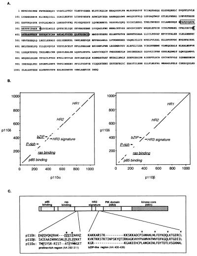
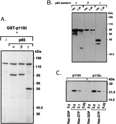
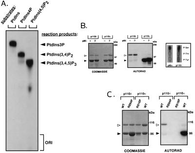
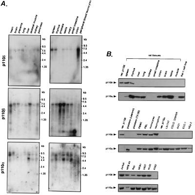
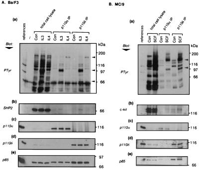
Phosphoinositide 3-kinases (PI3Ks) are a family of lipid kinases that have been implicated in signal transduction through tyrosine kinase- and heterotrimeric G-protein-linked receptors. We report herein the cloning and characterization of p110delta, a novel class I PI3K. Like p110alpha and p110beta, other class I PI3Ks, p110delta displays a broad phosphoinositide lipid substrate specificity and interacts with SH2/SH3 domain-containing p85 adaptor proteins and with GTP-bound Ras. In contrast to the widely distributed p110alpha and beta, p110delta is exclusively found in leukocytes. In these cells, p110alpha and delta both associate with the p85alpha and beta adaptor subunits and are similarly recruited to activated signaling complexes after treatment with the cytokines interleukin 3 and 4 and stem cell factor. Thus, these class I PI3Ks appear not to be distinguishable at the level of p85 adaptor selection or recruitment to activated receptor complexes. However, distinct biochemical and structural features of p110delta suggest divergent functional/regulatory capacities for this PI3K. Unlike p110alpha, p110delta does not phosphorylate p85 but instead harbors an intrinsic autophosphorylation capacity. In addition, the p110delta catalytic domain contains unique potential protein-protein interaction modules such as a Pro-rich region and a basic-region leucine-zipper (bZIP)-like domain. Possible selective functions of p110delta in white blood cells are discussed.


Figures

Scheme I
Scheme I
Figure 1
Figure 1
(A) Translated amino acid sequence of human p110δ cDNA. The Pro-rich region and the bZIP-like domain are indicated by open and shaded box, respectively. (B) Dot plot comparison of the full-length amino acid sequence of p110δ with that of p110α and β. Nonconserved sequence motifs are underlined. Dot plot comparisons were performed with the compare program (GCG package; ref. 39). (C) Comparison of the p110δ amino acid sequence flanking HR3 with the respective homologous regions of p110α and β. Amino acid numbering is that of p110δ. In the Pro-rich region, critical Pro enabling the formation of a left-handed polyPro type II helix in p110δ are indicated with an asterisk. In the bZIP-region, conserved Leu/Val/Ile residues of the leucine-zipper region are indicated with arrowheads.
Figure 2
Figure 2
Interaction of p110δ with p85 and Ras. (A) Insect cells were infected with baculovirus encoding GST–p110δ alone or in combination with viruses encoding either p85α, β, or γ. After 2 days, GST–p110δ was affinity-purified from the cell lysates with glutathione-Sepharose, washed, and analyzed by SDS/PAGE and Coomassie staining. (B) p110δ was immunoprecipitated from neutrophil cytosol and probed for the presence of different p85 isoforms by Western blotting. rec, Recombinant p85 purified from Sf9 cells. (C) GST–p110α/85α and GST–p110δ/85α (0.25 μg) were incubated with the indicated amount (in μg) of GTP- or GDP-loaded V12-Ras, washed, and probed for the presence of Ras by Western blotting as described (11, 12).
Figure 3
Figure 3
Enzymatic activity of p110δ. (A) In vitro lipid substrate specificity of p110δ. GST–p110δ/p85α was used in a lipid kinase assay with the indicated substrates in the presence of Mg2+. (B and C) Protein kinase activity of p110δ. Untagged p110α and δ [wild-type (WT) or kinase-defective mutants (p110α-R916P and p110δ-R894P)], in complex with p85α or β on platelet-derived growth factor receptor phosphopeptide beads, were subjected to an in vitro kinase reaction and further analyzed by SDS/PAGE, Coomassie staining, and autoradiography. Open and solid arrowheads point to p110 and p85 proteins, respectively. (B Right) Phosphoamino acid analysis of p85α and p110δ.
Figure 4
Figure 4
Tissue distribution of p110α, β, and δ. (A) Northern blot analysis of p110α, β, and δ expression. (B) Analysis of p110α and δ protein expression. Total cell lysate was loaded at 200 μg per lane. Platelets were lysed in either lysis buffer or in Laemmli gel loading buffer containing 2-mercaptoethanol. PMBC, peripheral blood mononuclear cell; PBL, peripheral blood lymphocyte.
Figure 5
Figure 5
Involvement of p110α and δ in cytokine signaling. Ba/F3 (A) and MC/9 (B) cell lines were stimulated with the indicated cytokines. Samples from control untreated cells are labeled Con. Total cell lysates and p110α and δ immunoprecipitates were separated by SDS/PAGE to prepare duplicate blots, the references for which were p110δ/85α (a, b, and d) or p110α/85α (c and e). Immunoblotting of naive blots was performed with 4G10 (anti-phosphotyrosine) (a) and anti-p110α (c). Blots were subsequently stripped and reprobed with anti-SHP2 (Ab), anti-kit (Bb), anti-p110δ (d), and anti-p85 antibodies (e). The arrowheads indicate the positions of p170 (IRS-2), p100, and p70 (SHP2) (Aa) and of p150 (c-kit) and p100 (Bb).

Similar articles

Cited by

References

    1. Zvelebil M J, MacDougall L, Leevers S, Volinia S, Vanhaesebroeck B, Gout I, Panayotou G, Domin J, Stein R, Koga H, Salim K, Linacre J, Das P, Panaretou C, Wetzker R, Waterfield M D. Philos Trans R Soc London. 1996;351:217–233. - PubMed
    1. Stephens L R, Jackson T R, Hawkins P T. Biochim Biophys Acta. 1993;1179:27–75. - PubMed
    1. Fry M. Biochim Biophys Acta. 1994;1226:237–268. - PubMed
    1. Kapeller R, Cantley L C. BioEssays. 1994;8:565–576. - PubMed
    1. Vanhaesebroeck B, Stein R, Waterfield M D. Cancer Surv. 1996;27:249–270. - PubMed

Publication types

MeSH terms

Associated data