. 1997 Apr 29;94(9):4406-11.
doi: 10.1073/pnas.94.9.4406.

Rescue of cardiac alpha-actin-deficient mice by enteric smooth muscle gamma-actin

Affiliations

Rescue of cardiac alpha-actin-deficient mice by enteric smooth muscle gamma-actin

A Kumar et al. Proc Natl Acad Sci U S A. .

Abstract

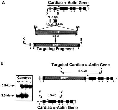
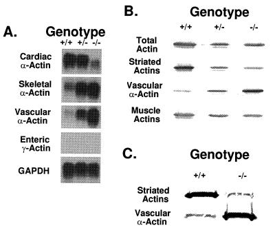
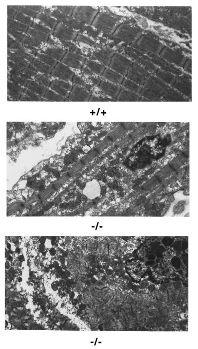
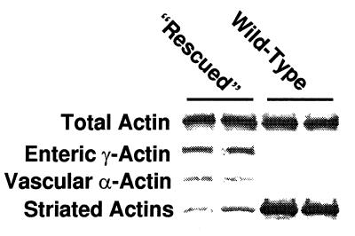
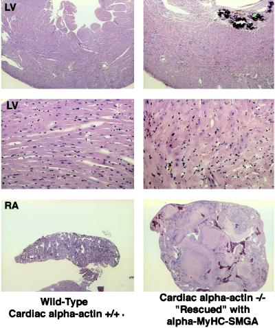
The muscle actins in higher vertebrates display highly conserved amino acid sequences, yet they show distinct expression patterns. Thus, cardiac alpha-actin, skeletal alpha-actin, vascular smooth muscle alpha-actin, and enteric smooth muscle gamma-actin comprise the major actins in their respective tissues. To assess the functional and developmental significance of cardiac alpha-actin, the murine (129/SvJ) cardiac alpha-actin gene was disrupted by homologous recombination. The majority ( approximately 56%) of the mice lacking cardiac alpha-actin do not survive to term, and the remainder generally die within 2 weeks of birth. Increased expression of vascular smooth muscle and skeletal alpha-actins is observed in the hearts of newborn homozygous mutants and also heterozygotes but apparently is insufficient to maintain myofibrillar integrity in the homozygous mutants. Mice lacking cardiac alpha-actin can be rescued to adulthood by the ectopic expression of enteric smooth muscle gamma-actin using the cardiac alpha-myosin heavy chain promoter. However, the hearts of such rescued cardiac alpha-actin-deficient mice are extremely hypodynamic, considerably enlarged, and hypertrophied. Furthermore, the transgenically expressed enteric smooth muscle gamma-actin reduces cardiac contractility in wild-type and heterozygous mice. These results demonstrate that alterations in actin composition in the fetal and adult heart are associated with severe structural and functional perturbations.


Figures

Figure 1
Figure 1
Targeted disruption of the cardiac α-actin gene. (A) Generation of the targeting fragment. Exon–intron organization of the cardiac α-actin gene is shown at the top. Open boxes represent noncoding exons, while dark boxes represent coding exons. Exons (–7) are numbered. The XbaI–SphI fragment used in the targeting construct includes 1.5 kb of sequence upstream and 0.7 kb of sequence downstream of the unique NaeI site. The NaeI site was converted to a SalI site, and the targeting construct was obtained by the ligation of a HPRT minigene SalI cassette. Since the HPRT minigene carries an XbaI site (not shown) the targeting fragment used for electroporation was released from the targeting construct by digestion with KpnI and SphI. The KpnI site is located in the vector (pUC 19). (B) Southern blot pattern of +/+, +/−, and −/− mice. Homologous recombination will lead to the generation of a 5.5-kb band, while the normal allele will give rise to a 3.5-kb band. The illustration to the right shows the location of the PvuII sites in the native and targeted gene. Bar indicates location of the probe used for Southern hybridization. K, KpnI; N, NaeI; S, SphI; Sa, SalI; V, PvuII; and X, XbaI.
Figure 2
Figure 2
Expression profiles of actin mRNA and protein from +/+, +/−, and −/− newborn mouse hearts. (A) Total heart RNA was analyzed for the expression of the different muscle actins. GAPDH was used as a control for the amount of RNA loaded. The minor band seen in the −/− heart includes transcripts deleted for sequences downstream of the HPRT insertion site (not shown). Note the increase in skeletal α-actin and vascular α-actin mRNA in the +/− and −/− hearts compared with the +/+ heart. Enteric SMGA expression is not observed in any of the three newborn hearts. (B) Total heart extracts were fractionated on an SDS/12% polyacrylamide gel and transferred to nitrocellulose. The actins were stained with different mAbs (C4 for total actin, 5C5 for the striated actins, 1A4 for vascular smooth muscle α-actin, and HUC 1–1 for muscle actin). Vascular smooth muscle α-actin shows an inverse correlation with cardiac α-actin gene dosage. The minor band seen in the −/− heart must represent skeletal α-actin, since mAb 5C5 recognizes both this isoform and cardiac α-actin. (C) Myofibrillar protein was similarly analyzed for its actin composition with the mAbs 1A4 and 5C5.
Figure 3
Figure 3
Electron micrographic analysis of newborn +/+ and −/− hearts. Note the myofibrillar disarray in the −/− heart. (×3,000.)
Figure 4
Figure 4
Extracts from adult hearts of +/+ and −/− rescued mice were electrophoresed on SDS/polyacrylamide gels and the actins were detected by staining with the different mAbs. The −/− rescued hearts have an abundance of enteric SMGA and minor amounts of vascular smooth muscle and striated (skeletal) actins. The total actin content is, however, similar in all the hearts.
Figure 5
Figure 5
Histopathology of a −/− rescued heart indicates hypertrophy. Comparison between Left and Right clearly shows myocyte hypertrophy in the left ventricle of the rescued heart. Also note the thrombi occluding a highly dilated right atrium (Bottom). LV, left ventricle; RA, right atrium. (Top ×15, Middle ×40, and Bottom ×4.)

Similar articles

Cited by

References

    1. Vandekerckhove J, Weber K. Differentiation. 1979;14:123–133. - PubMed
    1. Alonso S, Garner I, Vandekerckhove J, Buckingham M. J Mol Biol. 1990;211:727–738. - PubMed
    1. Vandekerckhove J, Bugaisky G, Buckingham M. J Biol Chem. 1986;261:1838–1843. - PubMed
    1. Ordahl C P. Dev Biol. 1986;117:488–492. - PubMed
    1. Minty A J, Alonso S, Caravatti M, Buckingham M E. Cell. 1982;30:185–192. - PubMed