

The nuclear matrix revealed by eluting chromatin from a cross-linked nucleus
- PMID: 9114009
- PMCID: PMC20742
- DOI: 10.1073/pnas.94.9.4446
The nuclear matrix revealed by eluting chromatin from a cross-linked nucleus
Abstract
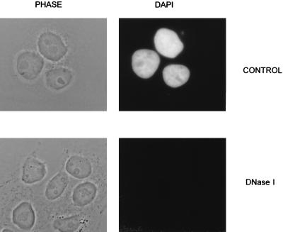
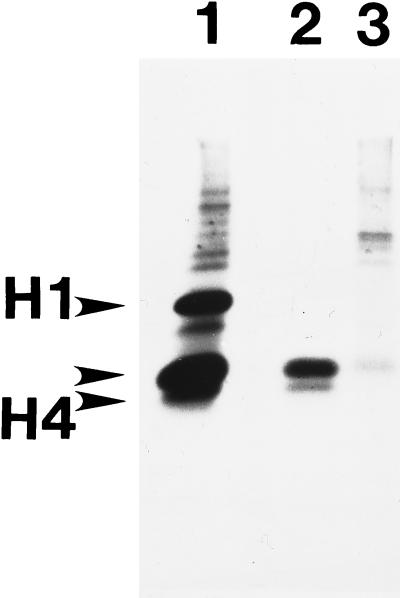
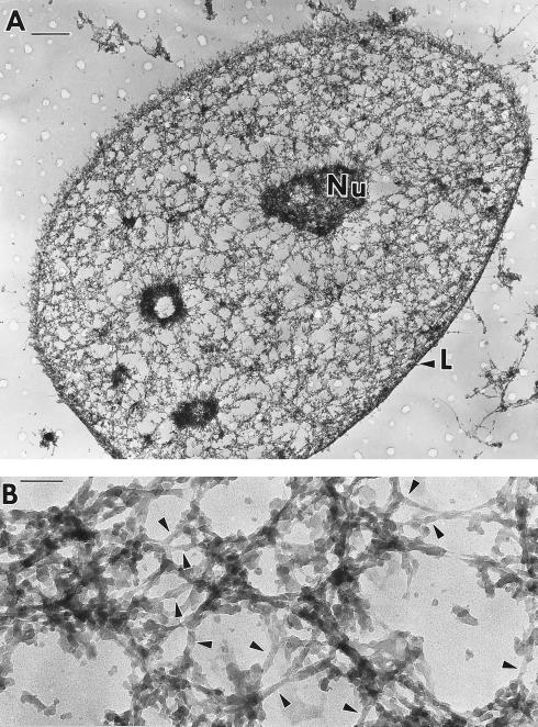
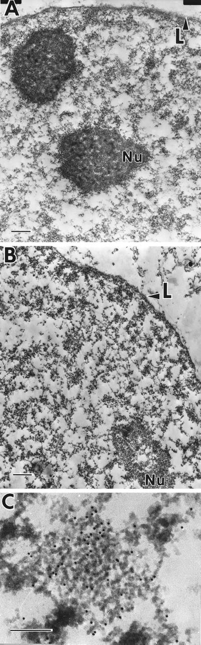
The nucleus is an intricately structured integration of many functional domains whose complex spatial organization is maintained by a nonchromatin scaffolding, the nuclear matrix. We report here a method for preparing the nuclear matrix with improved preservation of ultrastructure. After the removal of soluble proteins, the structures of the nucleus were extensively cross-linked with formaldehyde. Surprisingly, the chromatin could be efficiently removed by DNase I digestion leaving a well preserved nuclear matrix. The nuclear matrix uncovered by this procedure consisted of highly structured fibers, connected to the nuclear lamina and built on an underlying network of branched 10-nm core filaments. The relative ease with which chromatin and the nuclear matrix could be separated despite extensive prior cross-linking suggests that there are few attachment points between the two structures other than the connections at the bases of chromatin loops. This is an important clue for understanding chromatin organization in the nucleus.
Figures
Similar articles
-
The nuclear matrix prepared by amine modification.Proc Natl Acad Sci U S A. 1999 Feb 2;96(3):933-8. doi: 10.1073/pnas.96.3.933. Proc Natl Acad Sci U S A. 1999. PMID: 9927671 Free PMC article.
-
Core filaments of the nuclear matrix.J Cell Biol. 1990 Mar;110(3):569-80. doi: 10.1083/jcb.110.3.569. J Cell Biol. 1990. PMID: 2307700 Free PMC article.
-
Experimental observations of a nuclear matrix.J Cell Sci. 2001 Feb;114(Pt 3):463-74. doi: 10.1242/jcs.114.3.463. J Cell Sci. 2001. PMID: 11171316 Review.
-
The nonchromatin substructures of the nucleus: the ribonucleoprotein (RNP)-containing and RNP-depleted matrices analyzed by sequential fractionation and resinless section electron microscopy.J Cell Biol. 1986 May;102(5):1654-65. doi: 10.1083/jcb.102.5.1654. J Cell Biol. 1986. PMID: 3700470 Free PMC article.
-
Localization of nuclear matrix core filament proteins at interphase and mitosis.Cell Biol Int Rep. 1992 Aug;16(8):811-26. doi: 10.1016/s0309-1651(05)80024-4. Cell Biol Int Rep. 1992. PMID: 1446351 Review.
Cited by
-
NMPdb: Database of Nuclear Matrix Proteins.Nucleic Acids Res. 2005 Jan 1;33(Database issue):D160-3. doi: 10.1093/nar/gki132. Nucleic Acids Res. 2005. PMID: 15608168 Free PMC article.
-
The matrix attachment region in the Chinese hamster dihydrofolate reductase origin of replication may be required for local chromatid separation.Proc Natl Acad Sci U S A. 2003 Mar 18;100(6):3281-6. doi: 10.1073/pnas.0437791100. Epub 2003 Mar 10. Proc Natl Acad Sci U S A. 2003. PMID: 12629222 Free PMC article.
-
Use of HCA in subproteome-immunization and screening of hybridoma supernatants to define distinct antibody binding patterns.Methods. 2016 Mar 1;96:75-84. doi: 10.1016/j.ymeth.2015.10.021. Epub 2015 Oct 30. Methods. 2016. PMID: 26521976 Free PMC article.
-
Nucleoskeleton of early bovine embryos and differentiated somatic cells: an ultrastructural and immunocytochemical comparison.Histochem Cell Biol. 2004 Jun;121(6):441-51. doi: 10.1007/s00418-004-0655-3. Epub 2004 May 25. Histochem Cell Biol. 2004. PMID: 15221414
-
The nuclear matrix prepared by amine modification.Proc Natl Acad Sci U S A. 1999 Feb 2;96(3):933-8. doi: 10.1073/pnas.96.3.933. Proc Natl Acad Sci U S A. 1999. PMID: 9927671 Free PMC article.
References
Publication types
MeSH terms
Substances
Grants and funding
LinkOut - more resources
Full Text Sources
Other Literature Sources
