. 1997 Apr 29;94(9):4566-71.
doi: 10.1073/pnas.94.9.4566.

Synergism between hypersensitive sites confers long-range gene activation by the beta-globin locus control region

Affiliations

Synergism between hypersensitive sites confers long-range gene activation by the beta-globin locus control region

E H Bresnick et al. Proc Natl Acad Sci U S A. .

Abstract

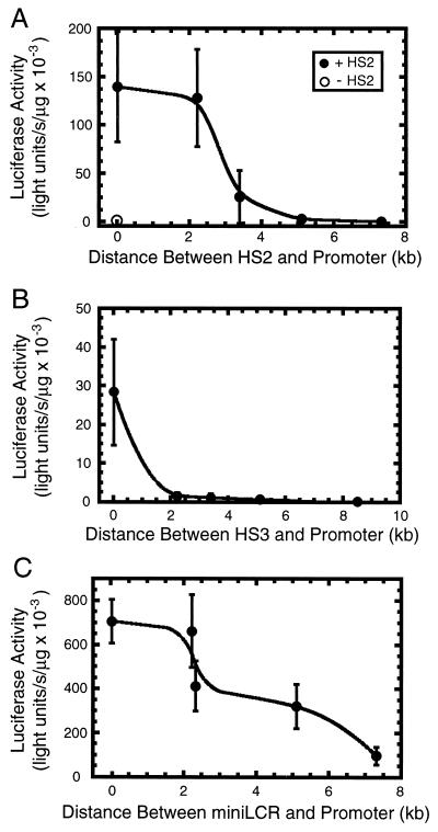
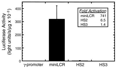
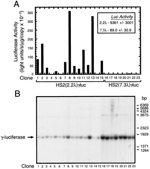
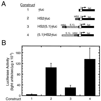
The human beta-globin locus control region (LCR) consists of four erythroid-specific DNaseI hypersensitive sites (HSs) at the 5' end of the beta-globin cluster. The LCR functions over a long distance on chromosome 11 to regulate transcription and replication of the beta-globin genes. To determine whether the HSs function independently or as an integrated unit, we analyzed the requirements for long-range transcriptional activation. If the HSs function independently, individual HSs would be expected to have long-range activity. In contrast, if long-range activity requires multiple HSs, individual HSs would have a limited functional distance. HS2, HS3, and a miniLCR containing multiple HSs, were separated from a gamma-globin promoter by fragments of phage lambda DNA. After stable transfection into K562 cells, HS2 had strong enhancer activity, but only when positioned close to the promoter. HS3 also had strong enhancer activity, although it was weaker than HS2 and more sensitive to the spacer DNA. The miniLCR had the strongest enhancer activity and functioned even at a distance of 7.3 kb. A model is proposed in which synergistic interactions between HSs confer long-range activation by creating a stable LCR nucleoprotein structure, which is competent for recruiting chromatin-modifying enzymes. These enzymes would mediate the well-characterized activity of the LCR to modulate chromatin structure.


Figures

Figure 1
Figure 1
Distance constraints for the transcriptional activation property of HS2, HS3, and the miniLCR. The three activating elements (A) HS2, (B) HS3, and (C) miniLCR, were subcloned upstream of a γ-globin promoter linked to a luciferase gene. Variable-length phage λ DNA fragments were subcloned between the activating element and promoter. The test constructs and a selection construct were linearized and cotransfected into K562 cells. Stably transfected pools of cells were isolated and assayed for luciferase activity as a measure of γ-globin promoter activity (mean ± SEM). The number of pools analyzed for the various constructs was: HS2γluc, 7; HS2(2.2)γluc, 7; HS2(3.4)γluc, 6; HS2(5.1)γluc, 7; HS2(7.3)γluc, 4; HS3γluc, 7; HS3(2.2)γluc, 8; HS3(3.4)γluc, 8; HS3(5.1)γluc, 5; HS3(8.5)γluc, 7; miniLCRγluc, 16; miniLCR(2.2)γluc, 12; miniLCR(2.3)γluc, 12; miniLCR(5.1)γluc, 12; miniLCR(7.3)γluc, 11. (A) The activity of a construct containing only the γ-globin promoter fused to the luciferase gene (pγluc) are shown by ○, whereas in AC the activity of constructs containing activating elements are shown by •. The luciferase activity of pγluc was 0.43 ± 0.49 light units per s/μg × 10−3 (n = 3).
Figure 2
Figure 2
Synergistic transcriptional activation by multiple hypersensitive sites. The luciferase values from Fig. 1, representing the activity of constructs containing a 5.1-kb phage λ insert between the activating element and promoter, are expressed in the graph. (Inset) The -fold activation of the test construct relative to the control plasmid containing the γ-globin promoter linked to the luciferase gene.
Figure 3
Figure 3
Low luciferase activity of cells containing pHS2(7.3λ)γluc is unrelated to template copy number. (A) The pHS2(2.2λ)γluc and pHS2(7.3λ)γluc constructs were linearized and cotransfected into K562 cells. Stably transfected clonal cell lines were isolated and assayed for luciferase activity as a measure of γ-globin promoter activity. (Inset) Mean luciferase activity of each set of clones [mean ± SEM, n = 15 and 8 for pHS2(2.2λ)γluc and pHS2(7.3λ)γluc constructs, respectively]. (B) Southern blot analysis of integrated templates. Genomic DNA was purified from the clonal cell lines of A and digested to completion with HindIII and XbaI to excise a fragment containing the γ-globin promoter linked to the luciferase gene. Genomic DNA (15 μg) was analyzed by Southern blot analysis with a luciferase probe.
Figure 4
Figure 4
Inhibition of transcriptional activation by placement of the 5.1-kb phage DNA fragment between HS2 and the promoter. (A) Structure of test constructs. (B) Test and selection constructs were linearized and cotransfected into K562 cells. Stably transfected pools of cells were isolated and assayed for luciferase activity as a measure of γ-globin promoter activity (mean ± SEM, n = 11, 9, and 9, and 9 for constructs 1–4, respectively).

References

    1. Felsenfeld G, Bresnick E H, Chung J, Reitman M. Molecular Biology of Hemoglobin Switching. Andover, MA: Intercept; 1995.
    1. Ptashne M. Nature (London) 1986;322:697–701. - PubMed
    1. Schleif R. Annu Rev Biochem. 1992;61:199–223. - PubMed
    1. Hager G L, Archer T K, Fragoso G, Bresnick E H, Tsukagoshi Y, John S, Smith C L. Cold Spring Harbor Symp Quant Biol. 1993;58:63–71. - PubMed
    1. Archer T K, Lefebvre P, Wolford R G, Hager G L. Science. 1992;255:1573–1576. - PubMed

Publication types

LinkOut - more resources